Posted on — Leave a comment

Kisspeptin-10 in Reproductive Neuroendocrine Research: GPR54/KISS1R Signaling and the HPG Axis

⚠️ For Research Purposes Only — The information in this article is provided strictly as a scientific reference for qualified laboratory researchers. Kisspeptin-10 and related compounds discussed here are not approved for human or veterinary use, are not drugs, and are not intended to diagnose, treat, cure, or prevent any disease. All experimental work should be performed only by trained personnel under appropriate institutional oversight.

Introduction

Few neuropeptides have rewritten a textbook as quickly as kisspeptin. In the space of roughly two decades, what began as the product of an obscure metastasis-suppressor gene (KISS1) has emerged as one of the most important upstream regulators of mammalian reproduction. Today, Kisspeptin-10 — a 10-residue C-terminal fragment of the parent peptide — is one of the most widely used research tools for probing the hypothalamic–pituitary–gonadal (HPG) axis in preclinical neuroendocrine studies.

This article is a technical primer for research laboratories working with Kisspeptin-10 in vitro and in animal models. It covers:

  • The molecular biology of the KISS1 gene and the KISS1R (GPR54) receptor
  • How Kisspeptin-10 interfaces with gonadotropin-releasing hormone (GnRH) neurons
  • Key preclinical findings that established kisspeptin as the “upstream regulator” of the HPG axis
  • Structural features that make the “-10” fragment the workhorse of laboratory research
  • Practical considerations for reconstitution, storage, and experimental design

Nothing in this article constitutes clinical or medical guidance. Kisspeptin-10 is discussed exclusively as a research reagent.

1. The KISS1 / KISS1R System: Molecular Background

The KISS1 gene was originally cloned in 1996 from the human melanoma cell line C8161 as a metastasis suppressor — hence “kiss” (a nod to Hershey, Pennsylvania’s “Kisses”). The primary translation product is a 145-amino acid precursor that is proteolytically processed into a 54-residue mature peptide, kisspeptin-54 (also known historically as metastin). Further cleavage yields shorter biologically active fragments sharing a common C-terminal decapeptide: Kisspeptin-10 (Tyr-Asn-Trp-Asn-Ser-Phe-Gly-Leu-Arg-Phe-NH₂).

All these fragments — KP-54, KP-14, KP-13, and KP-10 — act as full agonists at the same receptor: KISS1R, previously designated GPR54. KISS1R is a class A (rhodopsin-like) G protein-coupled receptor that signals predominantly through Gα_q/11, activating phospholipase C-β, generating IP₃ and diacylglycerol, mobilizing intracellular Ca²⁺, and activating protein kinase C and MAPK/ERK pathways.

The physiological relevance of this pathway was established when two independent groups in 2003 reported that loss-of-function mutations in KISS1R/GPR54 cause normosmic hypogonadotropic hypogonadism in humans and that Gpr54-null mice fail to enter puberty despite having morphologically normal GnRH neurons. Colledge’s 2004 review in Trends in Endocrinology and Metabolism consolidated these findings and established KISS1R as an obligate upstream gate for GnRH secretion (Colledge, 2004, DOI).

According to PubMed-indexed literature, kisspeptin signaling sits at the top of the reproductive neuroendocrine hierarchy: without functional KISS1R, the HPG axis cannot be activated, regardless of whether GnRH neurons, pituitary gonadotrophs, or gonads are intact.

2. Where Kisspeptin Neurons Live — and Why It Matters

In rodents, two major populations of kisspeptin-expressing neurons have been characterized:

  1. Arcuate nucleus (ARC) KNDy neurons — co-express kisspeptin, neurokinin B, and dynorphin. These are thought to constitute the “GnRH pulse generator,” providing the rhythmic excitatory drive that produces pulsatile GnRH release.
  2. Anteroventral periventricular nucleus (AVPV) neurons — sexually dimorphic (larger in females), implicated in the preovulatory LH surge in rodents.

In primates, the distribution is broadly similar, though the preoptic kisspeptin population is smaller and the ARC/infundibular population dominates pulse generation.

GnRH neurons themselves express KISS1R, and application of Kisspeptin-10 to GnRH neurons depolarizes them and triggers action potentials. This is the cellular basis for the axiom that “kisspeptin is the most potent known stimulator of GnRH secretion.”

3. Kisspeptin-10 vs. Full-Length Kisspeptin: Why the Fragment?

Herbison’s 2007 review of the genetics of puberty highlights why research has converged on shorter kisspeptin fragments (Herbison, 2007, DOI). The minimal bioactive core of the kisspeptin family lies in the C-terminal region, and structure-activity studies have shown that the C-terminal decapeptide (KP-10) retains essentially full intrinsic activity at KISS1R compared with the 54-residue parent.

Practical reasons KP-10 dominates preclinical research:

  • Synthetic accessibility: 10 residues are trivially produced by Fmoc solid-phase peptide synthesis, with high purity achievable on standard 0.1–0.25 mmol scales.
  • Defined pharmacology: KP-10 is a full KISS1R agonist in cellular assays with reported EC₅₀ values in the low nanomolar range.
  • Metabolic properties: KP-10 has a shorter plasma half-life than KP-54, which is useful when researchers want sharp on/off kinetics in bolus studies.
  • Reproducibility: Using a defined, single-sequence fragment reduces batch variability compared with recombinant or crudely processed parent material.

The trade-off is that KP-10 is cleared more rapidly than KP-54 in vivo, so in continuous-infusion or pulse-mimicking paradigms, researchers must account for the shorter effective duration.

4. Preclinical Evidence: What KP-10 Does to the HPG Axis

Animal models

Hameed and colleagues’ 2011 review in the Journal of Endocrinology summarizes the pre-2010 rodent and primate work: peripheral or central administration of Kisspeptin-10 produces robust, dose-dependent increases in luteinizing hormone (LH) and, to a lesser extent, follicle-stimulating hormone (FSH) via stimulation of hypothalamic GnRH neurons (Hameed et al., 2011, DOI). Chronic administration produces receptor desensitization in some paradigms, providing a useful tool for studying KISS1R tachyphylaxis.

Preclinical human volunteer research

Dhillo’s 2007 review in Reviews in Endocrine and Metabolic Disorders synthesized the first generation of preclinical volunteer research (Dhillo, 2007, DOI). In controlled experimental settings, intravenous administration of kisspeptin potently increased plasma LH, with smaller effects on FSH and downstream testosterone. Plasma kisspeptin was shown to be very low in non-pregnant adults but dramatically elevated during pregnancy, with the placenta identified as the source.

George et al. (2011, DOI) specifically investigated Kisspeptin-10 in an experimental volunteer setting and found that KP-10 is a potent stimulator of LH and increases LH pulse frequency — providing direct evidence that the decapeptide acts at the level of the GnRH pulse generator rather than the pituitary.

Abbara and colleagues (2018, DOI) reported that the hypothalamic response to kisspeptin-54 and the pituitary response to GnRH were preserved in healthy older men.

Kisspeptin antagonists as tools

Roseweir and Millar (2013, DOI) reviewed the development of peptide and small-molecule kisspeptin antagonists. These tools — used exclusively in preclinical research settings — have been invaluable for dissecting which aspects of reproductive physiology depend on endogenous KISS1R tone, including pubertal onset, estrous cyclicity, and the preovulatory LH surge.

All of the above is preclinical or experimental research data. None of it constitutes medical advice, a dosing recommendation, or an indication of safety or efficacy in any clinical population.

5. Structural Biology of Kisspeptin-10

Kisspeptin-10 is the C-terminal decapeptide of kisspeptin-54, with the sequence:

H-Tyr-Asn-Trp-Asn-Ser-Phe-Gly-Leu-Arg-Phe-NH₂

Important structural features relevant to laboratory handling:

  • Molecular formula: C₆₃H₈₃N₁₇O₁₄
  • Molecular weight: ~1302.5 Da (monoisotopic mass approximately 1301.63 Da)
  • C-terminal amidation: The C-terminal Phe is an amide, not a free carboxylic acid. This is critical for KISS1R binding; the free-acid analog shows markedly reduced potency.
  • RFamide motif: The C-terminal -Arg-Phe-NH₂ motif is shared with other RFamide family peptides (e.g., neuropeptide FF, prolactin-releasing peptide). Assay design should account for possible RFamide cross-reactivity.
  • Susceptible residues: Met is absent (good — no oxidation risk), but Trp is present and is vulnerable to photooxidation and some radical-generating conditions. Asn residues can undergo deamidation under high pH or prolonged storage in solution.

These chemical properties shape reconstitution and storage decisions (see Section 7).

6. Receptor Signaling: What the Assays Actually Measure

In vitro KISS1R assays commonly used in preclinical research include:

  • Intracellular Ca²⁺ mobilization (Fluo-4 or Fura-2): The most common readout, reflecting direct Gα_q activation. Typical time course: seconds to minutes after agonist addition.
  • Inositol phosphate (IP₁) accumulation: Homogeneous time-resolved fluorescence (HTRF) IP-One assays are widely used for GPCR profiling. Captures cumulative signaling over 30–60 minutes.
  • ERK1/2 phosphorylation: Measured by Western blot, AlphaLISA, or HTRF pERK assays. Captures downstream signaling with slower kinetics than Ca²⁺ (peak 5–15 min).
  • β-arrestin recruitment (e.g., PathHunter assays): Relevant for studying biased agonism at KISS1R and receptor desensitization.
  • Receptor internalization: BRET-based or immunofluorescence methods; relevant for tachyphylaxis studies.

Reported KP-10 EC₅₀ values in HEK293 or CHO cells heterologously expressing KISS1R typically fall in the 0.1–5 nM range, depending on assay format and receptor expression level. Researchers comparing analogs should always run a KP-10 reference curve on the same day to normalize between experiments.

Signaling bias considerations

KISS1R is primarily Gα_q/11-coupled, but like most class A GPCRs it can also recruit β-arrestin and engage Gα_i pathways in some systems. A peptide that activates Ca²⁺ signaling strongly but fails to recruit β-arrestin is said to be “Gq-biased”; the inverse is “arrestin-biased.” These distinctions matter for understanding why some kisspeptin analogs show different in vivo profiles despite identical Ca²⁺ EC₅₀ values. When designing structure–activity studies, including both Ca²⁺/IP₁ and β-arrestin readouts for each analog is the gold standard.

Species differences

KISS1R sequence conservation is high among mammals, but not perfect. Rodent KISS1R and human KISS1R differ in a small number of residues within the ligand-binding pocket, which can produce 2–10-fold potency differences for certain analogs. When running cross-species comparative studies, confirm the species origin of the receptor construct used in each assay.

6a. Structure-Activity Relationships of Kisspeptin-10 Analogs

The decapeptide scaffold of KP-10 has been extensively mined for structure-activity relationships in preclinical research. Key findings:

  • C-terminal amidation is essential: The free-acid analog (C-terminal -COOH instead of -CONH₂) shows dramatically reduced KISS1R potency. This is a common theme across RFamide peptides.
  • Arg9 is critical: Substitution of Arg9 with Ala or Lys reduces potency by 10- to 100-fold, reflecting its role in receptor engagement via electrostatic contacts.
  • Phe10 tolerates some substitution: Certain bulky aromatic analogs (e.g., 2-Nal, homophenylalanine) are tolerated, providing handles for affinity labels and photo-crosslinking studies.
  • N-terminal extension tolerated: Adding residues to the N-terminus (converting KP-10 into KP-11, KP-13, KP-14, KP-54) does not abolish activity; rather, it can modify pharmacokinetics and stability without disrupting KISS1R engagement.
  • D-amino acid substitutions at non-critical positions: Used to probe protease resistance for in vivo preclinical applications. Fully D-amino acid or retro-inverso analogs typically lose activity, but single D-substitutions at tolerant positions preserve it.

These SAR data underpin the rational design of kisspeptin antagonists, agonist analogs with altered pharmacokinetics, and photo-affinity probes for receptor mapping studies.

7. Practical Laboratory Considerations

Reconstitution

Kisspeptin-10 is reasonably soluble in aqueous buffers due to its polar residues (Asn, Ser) and basic C-terminal Arg. Suggested approaches for research use:

  • Primary stock: Dissolve lyophilized peptide in sterile research-grade water or dilute acetic acid (0.1% v/v) at 1 mg/mL. Mild acidification helps solubilize aggregates from the lyophilization process.
  • Working dilutions: Dilute further in assay buffer (e.g., HBSS + 0.1% BSA, or DMEM + 0.1% BSA). BSA is important to prevent non-specific adsorption of low-nanomolar working solutions to plastic.
  • Avoid: Prolonged incubation at high pH (>8) or repeated freeze–thaw of dilute stocks.

Storage

For lyophilized reference material:

  • Long-term: −20 °C or −80 °C, desiccated, protected from light.
  • Short-term: 2–8 °C is acceptable for a few weeks once sealed, provided the original container is kept desiccated.

For reconstituted working solutions:

  • Single-use aliquots: Prepare aliquots matched to your typical assay volume (e.g., 50 µL at 100 µM) so each tube is thawed once.
  • Freeze–thaw: Limit to ≤3 cycles; beyond that, expect measurable loss of potency due to Trp oxidation and Asn deamidation.

Stability assessment

If you are maintaining a stock over months, periodically re-check purity by RP-HPLC and identity by LC-MS. Signs of degradation include new HPLC peaks, broadening of the main peak, and the appearance of mass peaks +16 (oxidation) or −17 (deamidation + cyclization) relative to the intact peptide.

Experimental design notes

  • Time course: KP-10 acts rapidly in vitro (Ca²⁺ responses within seconds).
  • Desensitization: Repeated or prolonged exposure induces KISS1R desensitization; allow recovery time between stimulations in cell-based assays.
  • Controls: Always include a vehicle control matched to the peptide’s excipients (e.g., 0.1% BSA in saline) and, where possible, a scrambled-sequence peptide of the same length.
  • Concentration–response design: In Ca²⁺ or IP₁ assays, a typical concentration range is 0.01 nM to 1 µM in half-log steps, giving 8–10 points across the curve. This captures both the bottom plateau, the inflection, and the top plateau needed for accurate EC₅₀ fitting.
  • Non-specific binding: Include a BSA-only vehicle control and, where possible, a KISS1R-negative cell line (parental HEK293 or CHO without KISS1R transfection) to subtract background responses.

Animal study considerations

In rodent or larger-animal preclinical studies, additional considerations apply:

  • Blood sampling frequency: Because LH is pulsatile, frequent sampling (every 5–10 minutes for 1–2 hours) is required to accurately capture pulse frequency effects. Single time-point sampling misses the pulse dynamics that are often the most scientifically interesting readout.
  • Assay platform: LH can be measured by radioimmunoassay (legacy), ELISA (common), or LC-MS (emerging). Each has different sensitivity and dynamic range; choose based on the species and expected hormone concentrations.
  • Sex differences: Kisspeptin responses differ between males and females and between reproductive stages. Experimental design should control for or explicitly examine these variables.
  • Estrous/menstrual cycle stage: In cycling females, endogenous kisspeptin and GnRH tone vary dramatically; stage-matched comparisons are essential.

7a. Receptor Desensitization and Tachyphylaxis

One of the most studied features of KISS1R pharmacology in research settings is its capacity for rapid desensitization. Sustained exposure to kisspeptin — whether in vitro by prolonged incubation or in vivo by continuous infusion — produces a progressive decline in downstream signaling that is qualitatively distinct from simple receptor occupancy. This has both practical and scientific implications:

  • Practical: Continuous infusion paradigms produce smaller cumulative LH responses than pulsatile delivery, even when the total kisspeptin exposure is matched. Desensitization therefore has to be characterized before results from chronic-exposure models are interpreted.
  • Scientific: The mechanism of desensitization involves β-arrestin recruitment, receptor internalization, and potentially transcriptional downregulation of KISS1R expression. This makes KISS1R a useful model system for studying GPCR tachyphylaxis and the relationship between biased signaling and receptor trafficking.

For structure–activity studies of analogs intended as long-acting research tools, desensitization profiles should be characterized in parallel with acute potency. A compound that is equipotent to KP-10 in an acute Ca²⁺ assay but produces more sustained signaling without desensitization could be a more useful research tool for chronic studies — or, conversely, could produce confounding off-target effects via non-physiological tonic activation.

8. Frequently Asked Research Questions

Q1: What is the difference between Kisspeptin-10, Kisspeptin-54, and “metastin”?
Kisspeptin-54 is the mature cleavage product of the prepro-kisspeptin precursor. “Metastin” is an older name for the same peptide, derived from its original identification as a metastasis suppressor. Kisspeptin-10 is the C-terminal 10 amino acids of kisspeptin-54 and retains full intrinsic activity at KISS1R. For most in vitro mechanistic research, KP-10 is preferred; for longer-duration in vivo paradigms, KP-54 may be chosen for its longer plasma half-life.

Q2: Is Kisspeptin-10 selective for KISS1R?
Kisspeptin-10 is highly selective for KISS1R compared with most unrelated GPCRs, but it shares the RFamide C-terminal motif with other RFamide-family peptides. Cross-reactivity with NPFF receptors has been reported at high micromolar concentrations but is not a concern at the low-nanomolar concentrations used in typical KISS1R assays.

Q3: How should I handle Kisspeptin-10 to preserve activity?
Keep lyophilized material desiccated at −20 °C or −80 °C. Prepare single-use aliquots upon reconstitution. Include carrier protein (0.1% BSA) in dilute working solutions to prevent adsorption. Avoid repeated freeze–thaw and prolonged exposure to alkaline pH.

Q4: What vehicle should I use for in vitro KP-10 assays?
HEPES- or bicarbonate-buffered saline at physiological pH (7.2–7.4) with 0.1% BSA is a standard starting point. For Ca²⁺ imaging, HBSS with HEPES and BSA works well. Avoid buffers containing divalent cation chelators if you care about KISS1R coupling efficiency.

Q5: Why do reported potencies vary across papers?
Differences in KISS1R expression level, cellular background (HEK vs. CHO vs. primary neurons), assay readout (Ca²⁺ vs. IP₁ vs. ERK vs. β-arrestin), and peptide purity all contribute. Always normalize to a same-day reference curve, and report the assay conditions in full in your methods section.

References

  1. Colledge, W. H. (2004). GPR54 and puberty. Trends in Endocrinology and Metabolism, 15(9), 448–453. DOI: 10.1016/j.tem.2004.09.008 (PMID: 15519892)
  2. Dhillo, W. S. (2007). The neuroendocrine physiology of kisspeptin in the human. Reviews in Endocrine and Metabolic Disorders, 8(1), 41–46. DOI: 10.1007/s11154-007-9029-1 (PMID: 17323132)
  3. Herbison, A. E. (2007). Genetics of puberty. Hormone Research, 68 Suppl 5, 75–79. DOI: 10.1159/000110583 (PMID: 18174715)
  4. Hameed, S., Jayasena, C. N., & Dhillo, W. S. (2011). Kisspeptin and fertility. Journal of Endocrinology, 208(2), 97–105. DOI: 10.1677/JOE-10-0265 (PMID: 21084385)
  5. George, J. T., Veldhuis, J. D., Roseweir, A. K., et al. (2011). Kisspeptin-10 is a potent stimulator of LH and increases pulse frequency in men. Journal of Clinical Endocrinology & Metabolism, 96(8), E1228–E1236. DOI: 10.1210/jc.2011-0089 (PMID: 21632807)
  6. Roseweir, A. K., & Millar, R. P. (2013). Kisspeptin antagonists. Advances in Experimental Medicine and Biology, 784, 159–186. DOI: 10.1007/978-1-4614-6199-9_8 (PMID: 23550006)
  7. Abbara, A., Eng, P. C., Phylactou, M., et al. (2018). Hypothalamic response to Kisspeptin-54 and pituitary response to gonadotropin-releasing hormone are preserved in healthy older men. Neuroendocrinology, 107(3), 245–256. DOI: 10.1159/000488452 (PMID: 29544222)
  8. Sills, E. S., & Walsh, A. P. (2008). The GPR54-kisspeptin complex in reproductive biology: neuroendocrine significance and implications for ovulation induction and contraception. Neuroendocrinology Letters, 29(6), 846–851. (PMID: 19112386)
  9. Barbieri, R. L. (2014). The endocrinology of the menstrual cycle. Methods in Molecular Biology, 1154, 145–169. DOI: 10.1007/978-1-4939-0659-8_7 (PMID: 24782009)

Citations retrieved from PubMed. Please consult the original sources for full methodological detail.


Not for human consumption. For laboratory research only.

Disclaimer: All products sold by CertaPeptides are intended for research purposes only. Not for human or veterinary use. Not for consumption. Nothing in this article is medical advice, and no claims of safety, efficacy, diagnosis, treatment, prevention, or cure are made for Kisspeptin-10 or any related compound. Researchers are responsible for complying with all applicable laws, institutional policies, and ethical guidelines governing the handling of research peptides.
Posted on — Leave a comment

MOTS-c: A Research Guide to the Mitochondrial-Derived Peptide Behind Metabolic and Aging Studies

⚠️ For Research Purposes Only — This article discusses MOTS-c strictly as a laboratory research compound. It is not a drug recommendation, dosage guide, or medical advice. MOTS-c is not for human consumption. It is not intended to diagnose, treat, cure, or prevent any disease. All claims below refer to published preclinical research, in vitro investigations, or peer-reviewed literature cited for scientific context only.

Introduction

MOTS-c (mitochondrial open reading frame of the 12S rRNA-c) is a short bioactive peptide encoded within the mitochondrial genome rather than the nuclear genome. Its discovery, first formally described in 2015 [1], represented a conceptual shift in mitochondrial biology: for decades, mitochondria were thought to produce only 13 proteins from their own DNA, all integral to the electron transport chain. MOTS-c and its sibling peptides (humanin, the SHLP family) expanded that count and introduced the concept of mitochondrial-derived peptides (MDPs) acting as signaling molecules, not just structural or catalytic components of the respiratory machinery [2][3].

For researchers interested in mitochondrial biology and metabolic signaling, MOTS-c is scientifically compelling because it functions as a retrograde signal — a message from mitochondria back to the nucleus and to distant tissues — in response to metabolic stress. This article reviews the published evidence on MOTS-c’s structure, mechanism of action, and the principal research areas in which it has been investigated.

Molecular Structure and Biochemistry

MOTS-c is a 16-amino-acid peptide with the published sequence Met-Arg-Trp-Gln-Glu-Met-Gly-Tyr-Ile-Phe-Tyr-Pro-Arg-Lys-Leu-Arg. It is encoded by a short open reading frame (sORF) embedded within the mitochondrial 12S rRNA gene, on the opposite strand from the canonical rRNA transcript. The peptide is translated on cytoplasmic ribosomes from a mitochondrially-derived RNA template, a feature that distinguishes it from mitochondrially-translated respiratory chain subunits [1].

Because the mitochondrial genetic code differs slightly from the nuclear code, the MOTS-c sORF would encode a different sequence if read inside the mitochondrial matrix. The fact that MOTS-c is translated cytoplasmically — using the standard genetic code — is an important mechanistic point for researchers, and it has implications for how synthetic MOTS-c is designed and validated against endogenous species.

Key biochemical features relevant to laboratory research:

  • Small size (~1.9 kDa) makes MOTS-c suitable for synthesis by standard solid-phase Fmoc peptide chemistry without complex folding requirements.
  • Basic character (two arginines, one lysine) gives the peptide a net positive charge at physiological pH and contributes to its interaction with acidic subcellular compartments and nucleic acids.
  • Evolutionary conservation: Comparative genomics research has demonstrated synonymous codon bias and high N-terminal sequence conservation across vertebrates, suggesting that MOTS-c and humanin are under natural selection — a strong argument for biological functionality [4].
  • Subcellular distribution: MOTS-c is found in mitochondria, cytoplasm, and, under metabolic stress, in the nucleus where it engages stress-responsive transcription factors [5].
  • Circulating peptide: MOTS-c is detectable in plasma in humans and rodents, and circulating levels decline with age in published research cohorts [2][6].

Mechanism of Action in Research Models

MOTS-c operates through at least two mechanistically distinct modes described in the published research literature:

1. AMPK activation and folate-cycle modulation. The foundational 2015 research from the Cohen laboratory reported that MOTS-c treatment activates AMP-activated protein kinase (AMPK) in cultured cells and in skeletal muscle. The mechanism involves inhibition of the folate cycle and the tethered de novo purine biosynthesis pathway, producing an accumulation of AICAR-like intermediates that activate AMPK [1]. Downstream of AMPK activation, MOTS-c treatment was reported to influence glucose uptake in skeletal muscle in research mice.

2. Retrograde signaling to the nucleus. A 2018 follow-up study demonstrated that under metabolic stress (glucose restriction), MOTS-c translocates from mitochondria and cytoplasm into the nucleus, where it engages the antioxidant response element (ARE) transcriptional program and interacts with stress-responsive transcription factors including NFE2L2/NRF2 [5]. This was the first direct evidence that a peptide encoded in the mitochondrial genome could regulate nuclear gene expression, providing a molecular basis for “mitonuclear communication.”

A 2023 study extended the NRF2 interaction story to dopaminergic neuron research models, reporting that exogenous MOTS-c activated the Nrf2/HO-1/NQO1 antioxidant axis in PC12 cells and in rat striatum exposed to rotenone, a mitochondrial complex I inhibitor used as a research model of dopaminergic neurotoxicity [7].

Additional mechanistic threads in the published literature include:

  • Adipose thermogenesis. MOTS-c treatment in cold-exposed mice upregulated brown adipose tissue thermogenic gene expression and promoted “browning” of white adipose tissue, via ERK signaling [8].
  • Skeletal muscle and myostatin. Research in differentiated C2C12 myotubes and diet-induced obese mice reported that MOTS-c reduces myostatin expression and atrophy signaling via a CK2-PTEN-mTORC2-AKT-FOXO1 pathway [9].
  • Ferroptosis suppression. A 2023 study in myocardial ischemia-reperfusion-induced acute lung injury described MOTS-c as a suppressor of ferroptosis via the PPARγ signaling pathway in preclinical rat and cell culture models [10].

Key Research Areas

1. Metabolic Signaling Research

The metabolic research area is the oldest and most thoroughly characterized for MOTS-c. The 2015 discovery paper [1] established the baseline findings, identifying skeletal muscle as the peptide’s primary target organ with cellular action through AMPK activation and downstream effects on glucose handling.

Subsequent research has examined MOTS-c signaling across a range of cellular and animal models, including studies of its molecular interactions in other tissue contexts. A 2024 research report described MOTS-c interacting with LARS1 and the USP7 deubiquitinase in preclinical models [12], an example of how a metabolic signaling peptide can cross into other research domains when its molecular targets are present in multiple cell types.

For researchers designing metabolic experiments, MOTS-c offers a useful counterpoint to insulin or metformin as an AMPK-activating probe, because its pathway of activation is mechanistically distinct (via folate-cycle intermediates rather than direct allosteric binding).

2. Aging Biology Research

MOTS-c has become a subject of significant interest in aging research because: (a) circulating MOTS-c levels decline with age in human cohorts [2][6]; (b) mitochondrial dysfunction is a central “hallmark of aging” [6]; and (c) exogenous MOTS-c administration alters several age-associated metabolic phenotypes in research mice [1].

A 2022 review catalogued the published evidence for MOTS-c’s expression and signaling in aging research models, framing MOTS-c as both a biomarker and a research probe [6]. A 2023 translational review from Frontiers in Endocrinology surveyed MOTS-c’s expression across tissues, its decline with age, and the emerging possibility of synthetic-biology approaches to modulate its expression in research settings [13].

For researchers in the aging field, MOTS-c is scientifically useful as a dual readout: circulating levels can serve as a biomarker of mitochondrial secretory function, while exogenous administration provides an interventional probe of the mitonuclear communication axis [5][6].

3. Skeletal Muscle Signaling Research

Skeletal muscle signaling is a developing research area for MOTS-c. The 2016 free-radical-biology review from Lee et al. discussed MOTS-c in the context of muscle and fat metabolism, noting that its primary target organ (skeletal muscle) is a tissue in which AMPK signaling is a central mediator of metabolic adaptation [3]. Subsequent research has reported MOTS-c-dependent effects on muscle gene expression in preclinical models.

The myostatin work [9] is relevant to this research area at the molecular level: research in differentiated myotubes and diet-induced obese mice reported that MOTS-c modulates myostatin expression via the CK2/PTEN/AKT/FOXO1 axis, providing a mechanistic handle for researchers interested in linking mitochondrial stress signals to muscle protein turnover.

Stability, Storage, and Handling in the Laboratory

MOTS-c is a relatively straightforward peptide from a handling standpoint, but researchers should be aware of several practical points:

  • Lyophilized form: As supplied, MOTS-c is typically a white lyophilized powder. Long-term storage at -20°C or -80°C in a desiccated environment is standard research practice. Vials should be equilibrated to room temperature before opening to prevent atmospheric moisture condensation.
  • Reconstitution: Sterile water or 0.1% acetic acid are common reconstitution solvents for MOTS-c in research protocols. Its basic residues give it reasonable aqueous solubility; some researchers use dilute acetic acid for initial dissolution followed by buffer dilution.
  • pH sensitivity: Like most peptides with tryptophan and methionine residues (MOTS-c has one Trp and two Met), prolonged exposure to oxidizing conditions should be avoided. Degassed buffers and amber vials are recommended for quantitative research work.
  • Aliquoting: Repeated freeze-thaw cycles accelerate degradation. Best practice is to reconstitute once, aliquot into single-use volumes, and store frozen. Working dilutions should be made fresh on the day of experiment.
  • Characterization: HPLC and mass spectrometry confirmation is recommended for research-grade material, particularly given the similarity of MOTS-c to peptide fragments that may arise from synthesis byproducts.

Research Considerations and Limitations

Mixed literature on receptors. Unlike many peptide hormones, MOTS-c does not have a clearly defined single cell-surface receptor in the published literature. Its intracellular mechanism via AMPK activation and nuclear translocation is well supported, but the mechanism of cellular uptake — and whether it involves specific transporters or nonspecific endocytosis — remains an active area of research investigation.

Species and tissue specificity. Most mechanistic research has been conducted in rodent or human cell-line systems. Translational extrapolation to other species or to tissues with low MOTS-c expression requires careful interpretation.

Assay variability for endogenous MOTS-c. Because MOTS-c is small, highly basic, and present in low concentrations in plasma, quantification relies on immunoassays whose performance has varied across published research reports. Researchers designing biomarker studies should validate their assay with recombinant or synthetic standards.

Synthetic product heterogeneity. MOTS-c is available from multiple research suppliers at varying purities. For quantitative research work, HPLC purity >95% and mass-spectrometric confirmation of the full-length sequence are minimum expectations.

Limited human research. The vast majority of published MOTS-c research is preclinical (in vitro or rodent). Human data is largely observational (circulating levels as a biomarker), with controlled interventional studies remaining an open research frontier.

Frequently Asked Research Questions

Q1: Is MOTS-c a hormone, a signaling peptide, or an intracellular regulator?
Published research has characterized MOTS-c in all three modes. It is detectable in circulation (hormone-like), it acts on distant target tissues such as skeletal muscle (paracrine/endocrine signaling), and it translocates to the nucleus to regulate gene expression (intracellular regulator) [1][5][6]. Researchers should specify which mode they are studying in any given experiment.

Q2: How does MOTS-c differ from humanin, the other major mitochondrial-derived peptide?
Both MOTS-c and humanin are encoded by short open reading frames within the mitochondrial 12S and 16S rRNA regions, respectively. However, humanin primarily exerts anti-apoptotic and neuroprotective effects via cell-surface receptors (FPR2/3, CNTFR/WSX-1/gp130), while MOTS-c acts predominantly through intracellular AMPK activation and nuclear translocation [3][4]. Their functional repertoires overlap at the level of metabolic and stress-response signaling but differ mechanistically.

Q3: What is the evidence that MOTS-c is a genuine bioactive peptide rather than a translational curiosity?
The strongest evidence comes from (a) the reproducibility of MOTS-c effects across multiple independent laboratories since 2015 [1][5][11], (b) the evolutionary conservation of its coding sequence under synonymous codon bias analysis [4], and (c) the biological phenotypes produced by synthetic MOTS-c in defined preclinical models. Skepticism remains appropriate for any specific claim, but the cumulative evidence supports MOTS-c as a functional MDP.

Q4: What are the standard positive controls for MOTS-c experiments?
AMPK activation experiments typically use AICAR or metformin as positive controls [1]. Nuclear translocation studies use oxidative stressors such as tert-butyl hydroperoxide or glucose restriction as positive controls for the NRF2/ARE pathway [5]. For exercise-physiology research, acute exercise itself is a natural positive control.

Q5: Can MOTS-c be measured in clinical plasma samples?
Yes, several published research studies have reported circulating MOTS-c measurements in human cohorts using ELISA-based assays [2][6]. However, assay variability and small sample volumes remain technical challenges. Researchers intending to use MOTS-c as a biomarker should validate their assay against synthetic peptide standards and report recovery, linearity, and inter-assay variability.

References

According to PubMed, the following peer-reviewed articles were used as primary sources for this research overview. DOI links are provided where available.

  1. Lee C, Zeng J, Drew BG, et al. The mitochondrial-derived peptide MOTS-c promotes metabolic homeostasis and reduces obesity and insulin resistance. Cell Metab. 2015;21(3):443-54. PMID: 25738459. DOI
  2. Cobb LJ, Lee C, Xiao J, et al. Naturally occurring mitochondrial-derived peptides are age-dependent regulators of apoptosis, insulin sensitivity, and inflammatory markers. Aging (Albany NY). 2016;8(4):796-809. PMID: 27070352. DOI
  3. Lee C, Kim KH, Cohen P. MOTS-c: A novel mitochondrial-derived peptide regulating muscle and fat metabolism. Free Radic Biol Med. 2016;100:182-187. PMID: 27216708. DOI
  4. Gruschus JM, Morris DL, Tjandra N. Evidence of natural selection in the mitochondrial-derived peptides humanin and SHLP6. Sci Rep. 2023;13(1):14110. PMID: 37644144. DOI
  5. Kim KH, Son JM, Benayoun BA, Lee C. The Mitochondrial-Encoded Peptide MOTS-c Translocates to the Nucleus to Regulate Nuclear Gene Expression in Response to Metabolic Stress. Cell Metab. 2018;28(3):516-524.e7. PMID: 29983246. DOI
  6. Mohtashami Z, Singh MK, Salimiaghdam N, Ozgul M, Kenney MC. MOTS-c, the Most Recent Mitochondrial Derived Peptide in Human Aging and Age-Related Diseases. Int J Mol Sci. 2022;23(19):11991. PMID: 36233287. DOI
  7. Xiao J, Zhang Q, Shan Y, et al. The Mitochondrial-Derived Peptide (MOTS-c) Interacted with Nrf2 to Defend the Antioxidant System to Protect Dopaminergic Neurons Against Rotenone Exposure. Mol Neurobiol. 2023;60(10):5915-5930. PMID: 37380822. DOI
  8. Lu H, Tang S, Xue C, et al. Mitochondrial-Derived Peptide MOTS-c Increases Adipose Thermogenic Activation to Promote Cold Adaptation. Int J Mol Sci. 2019;20(10):2456. PMID: 31109005. DOI
  9. Kumagai H, Coelho AR, Wan J, et al. MOTS-c reduces myostatin and muscle atrophy signaling. Am J Physiol Endocrinol Metab. 2021;320(4):E680-E690. PMID: 33554779. DOI
  10. Lu P, Li X, Li B, et al. The mitochondrial-derived peptide MOTS-c suppresses ferroptosis and alleviates acute lung injury induced by myocardial ischemia reperfusion via PPARγ signaling pathway. Eur J Pharmacol. 2023;953:175835. PMID: 37290680. DOI
  11. Yin Y, Pan Y, He J, et al. The mitochondrial-derived peptide MOTS-c relieves hyperglycemia and insulin resistance in gestational diabetes mellitus. Pharmacol Res. 2021;175:105987. PMID: 34798268. DOI
  12. Yin Y, Li Y, Ma B, et al. Mitochondrial-Derived Peptide MOTS-c Suppresses Ovarian Cancer Progression by Attenuating USP7-Mediated LARS1 Deubiquitination. Adv Sci (Weinh). 2024;11(43):e2405620. PMID: 39321430. DOI
  13. Zheng Y, Wei Z, Wang T. MOTS-c: A promising mitochondrial-derived peptide for therapeutic exploitation. Front Endocrinol (Lausanne). 2023;14:1120533. PMID: 36761202. DOI

Disclaimer: All products sold by CertaPeptides are intended for research purposes only. Not for human or veterinary use. Not for consumption. This article is provided for scientific and educational purposes and does not constitute medical advice, dosing guidance, or an endorsement of any particular use. Researchers are responsible for complying with all applicable institutional, national, and international regulations governing the acquisition, handling, and study of peptide research compounds.

Posted on — Leave a comment

Peptide Lyophilization: The Freeze-Drying Science Behind Stable Research Peptides

⚠️ For Research Purposes Only — This article is a technical reference for qualified laboratory researchers working with peptide reagents. It discusses formulation and stability science, not therapeutic use. Peptides referenced as examples are not approved for human or veterinary use and are supplied strictly for laboratory research.

Introduction

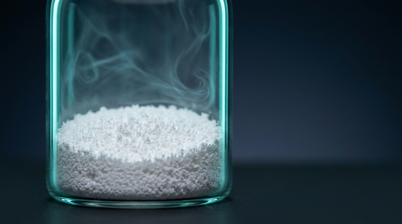
Walk into any peptide research lab and you’ll find a shelf full of small glass vials containing what looks like fluffy white residue, spun-glass thread, or a thin compact disk of solid. That solid is the whole reason the peptide inside is still usable months or years after synthesis. Lyophilization — freeze-drying — is the enabling technology of modern peptide distribution. It transforms an unstable aqueous solution into a dry, storage-stable cake that can survive shipping, cold-chain interruptions, and long-term freezer storage.

This article is a technical primer for research laboratories on how lyophilization actually works, why certain excipients are chosen, and how to interpret what you see when you open the vial. It covers:

  • The thermodynamics and physics of freeze-drying
  • The three stages of a standard lyophilization cycle
  • Cryoprotectants and lyoprotectants: what they do and why they matter
  • Cake structure, collapse temperature, and glass transitions
  • Residual moisture and its relationship to stability
  • Common failure modes — shrinkage, meltback, and collapse — and what they tell you

Nothing in this article is medical guidance. All discussion is in the context of research-use materials.

1. Why Lyophilize Peptides at All?

In solution, peptides are exposed to every degradation pathway their amino acid composition allows:

  • Hydrolysis of peptide bonds, especially at Asp-Pro and Asp-Xaa linkages.
  • Deamidation of Asn and (more slowly) Gln via succinimide intermediates.
  • Oxidation of Met, Cys, Trp, Tyr, and His, accelerated by dissolved oxygen, trace metals, and light.
  • Aggregation via non-covalent association, disulfide scrambling, or hydrophobic partitioning.
  • Physical instability — precipitation, gelation, adsorption to surfaces.

Drying the peptide removes water, which is the solvent for most of these reactions and the kinetic enabler of molecular motion. A properly lyophilized peptide stored cold and dry can remain chemically stable for years; the same peptide in solution at 4 °C may lose significant potency within weeks.

Izutsu (2018, DOI) provides a comprehensive overview of how freeze-drying is applied to pharmaceutical formulations — peptides, proteins, and small molecules alike (PMID: 30288720). The principles map directly onto research-grade peptide manufacturing.

2. Thermodynamics: What Actually Happens in a Freeze-Dryer

Lyophilization exploits a simple phase-diagram trick. At atmospheric pressure, water transitions ice → liquid → vapor. At pressures below the triple point of water (611 Pa, or 4.58 Torr), liquid water cannot exist — ice sublimes directly to vapor. A freeze-dryer operates in this regime: the product is frozen solid, the chamber is evacuated below the triple point, and heat is applied carefully to drive sublimation without melting.

The two critical temperatures that govern every cycle:

  • T′_g (glass transition of the maximally freeze-concentrated solute): The temperature below which the amorphous phase is a rigid glass. For sucrose–water systems, T′_g is approximately −32 °C; for trehalose, approximately −29 °C.
  • T_c (collapse temperature): The temperature at or above which the dried matrix loses enough mechanical integrity to collapse under vacuum. T_c is typically slightly above T′_g for amorphous systems.

Primary drying must be performed with the product temperature held below T_c — otherwise the cake collapses, trapping water and destroying the porous structure that enables fast reconstitution. This single constraint drives almost every design decision in a lyophilization cycle.

3. The Three Stages of a Lyophilization Cycle

Stage 1 — Freezing

The peptide solution is cooled in a controlled ramp, typically −0.5 to −1 °C/min, down to a shelf temperature of −40 to −50 °C. As ice nucleates, solutes are concentrated into the interstitial amorphous phase. The freezing step determines ice crystal size and, therefore, the geometry of pores through which water vapor must escape in the next stage.

Key variables:

  • Nucleation temperature: Spontaneous nucleation is stochastic and can occur anywhere from −8 to −18 °C, leading to batch variability. Controlled nucleation techniques (e.g., depressurization, ice fog seeding) produce more uniform cakes.
  • Cooling rate: Slow cooling produces larger ice crystals (bigger pores, faster sublimation) but can concentrate solutes and damage some peptides. Fast cooling produces finer crystals and finer pores, slowing primary drying.
  • Annealing: Holding the frozen product at a temperature above the glass transition (e.g., −15 °C) for 1–4 hours allows ice crystal ripening, producing larger, more uniform crystals and improving downstream drying efficiency.

Abdelwahed et al. (2006, DOI) characterized the impact of annealing on both primary and secondary drying kinetics in pharmaceutical formulations containing cryoprotectants such as sucrose and polyvinylpyrrolidone (PMID: 16904277). Their work showed that annealing can accelerate sublimation without compromising reconstituted product properties — one of the classic process-optimization findings in the field.

Stage 2 — Primary Drying (Sublimation)

Chamber pressure is reduced below the triple point of water (typically 50–200 mTorr), and shelf temperature is raised to supply latent heat of sublimation while keeping the product temperature below T_c. Ice sublimes from the surface of the frozen matrix downward and outward, leaving behind the amorphous solute phase laced with empty pores.

This is the longest stage of a typical cycle — often 20–80 hours, depending on product volume, vial geometry, and cake design. Shelf temperature is the driver; chamber pressure fine-tunes heat transfer. Product temperature is the constrained variable.

Researchers designing a cycle monitor product temperature with thermocouples in representative vials. If the product exceeds T_c, cake collapse begins and the cycle must be aborted or recovered.

Stage 3 — Secondary Drying (Desorption)

After sublimation is complete, roughly 5–20% water remains bound to the solute matrix. Secondary drying raises the shelf temperature to 20–40 °C at reduced pressure to drive off this residual bound water by desorption. Duration is typically 5–15 hours.

The endpoint of secondary drying is specified as a residual moisture target — commonly 1–3% w/w for most peptide products, though the exact target is determined by stability data for the specific molecule. Karl Fischer titration is the gold standard for residual moisture measurement in research and manufacturing.

4. Excipients: Cryoprotectants, Lyoprotectants, Bulking Agents

A lyophilized peptide formulation is almost never just peptide and water. Excipients do four main jobs:

Cryoprotectants (protection during freezing)

Sugars and polyols form hydrogen bonds that stabilize the peptide’s native conformation when water is removed. Sucrose and trehalose are the dominant cryoprotectants in peptide and protein lyophilization because:

  • They are amorphous glass-formers at typical process temperatures.
  • They have relatively high T′_g values (~−32 °C and ~−29 °C respectively), allowing warmer primary drying and shorter cycles.
  • They form direct hydrogen bonds with peptide backbone amides, substituting for the water-bridge network that normally stabilizes peptide structure (the “water replacement hypothesis”).
  • They vitrify into a high-viscosity glass that mechanically immobilizes the peptide and kinetically arrests degradation reactions (the “vitrification hypothesis”).

Trehalose is often preferred for more labile peptides because it does not contain a reducing group and cannot undergo Maillard reactions with lysine side chains. Sucrose is cheaper but can hydrolyze under acidic conditions to release reducing sugars that react with Lys and Arg.

Li et al. (1996, DOI) demonstrated in a landmark study that reducing sugars can accelerate chemical degradation of lyophilized peptides through Maillard-type reactions (PMID: 8863280). This paper is often cited as the definitive case for using non-reducing disaccharides with Lys-rich sequences.

Lyoprotectants (protection during drying and storage)

Many cryoprotectants also serve as lyoprotectants. The distinction is useful: cryoprotection addresses damage during freezing (osmotic, ice-interface), while lyoprotection addresses damage during water removal and subsequent storage. Sucrose, trehalose, and some amino acids (glycine, arginine, histidine) fall into both categories.

Bulking agents

When the peptide concentration is very low (as is common for potent research peptides at milligram scale), the solute mass is not enough to form a mechanically coherent cake. Mannitol, glycine, and sometimes lactose are added as bulking agents. Mannitol crystallizes during freezing, producing a rigid crystalline scaffold — but this can be problematic because crystalline bulking agents do not contribute to lyoprotection of the amorphous peptide phase. A common compromise is a mannitol:sucrose 3:1 or 4:1 blend: mannitol for cake structure, sucrose for lyoprotection.

Buffers and pH modifiers

Peptide stability is strongly pH-dependent. Common buffers include phosphate, acetate, histidine, and citrate. Phosphate can crystallize during freezing, causing dramatic pH shifts (up to 3 pH units) as one phosphate species crystallizes preferentially — a classic failure mode that damages pH-sensitive peptides.

Surfactants

Low-concentration polysorbate 20 or 80 (0.001–0.01% w/v) is often added to protect peptides from air–liquid interface denaturation during filling and reconstitution.

Acidifiers

Some peptides (e.g., those reconstituted in acetic acid) include acetate buffer in the lyophilized cake. Volatile acidifiers can partially evaporate during drying, shifting the final cake pH.

5. Cake Structure and What It Tells You

A properly lyophilized cake should have:

  • Full volume: The cake should occupy the same volume as the pre-lyophilization fill. Shrinkage suggests collapse during drying.
  • Uniform appearance: Fine-textured, typically white, evenly distributed from top to bottom of the vial.
  • Mechanical integrity: The cake should not crumble when the vial is gently tapped; nor should it stick as a hard glass.
  • Rapid reconstitution: Water added to a well-formed cake should produce a clear solution within seconds to minutes, depending on concentration.

Failure modes to recognize:

  • Collapse: The cake has sunk to a fraction of its original volume, often with a glassy or shrunken appearance. Caused by exceeding T_c during primary drying. Reconstitution time is longer and product may be partially denatured.
  • Meltback: A wet layer at the bottom of the vial, caused by reaching the melting point of the concentrated amorphous phase. The product may still reconstitute but has likely lost some stability.
  • Powdering / shattering: The cake has broken apart during handling. Can indicate low residual moisture brittleness or mechanical damage.
  • Discoloration: Yellow or brown tint suggests Maillard browning (reducing sugar + Lys/Arg) or oxidation.
  • Fiber-like strands: Normal for some highly amorphous formulations; not necessarily a failure.

6. Residual Moisture: The Critical Spec

Residual moisture is one of the most important quality attributes of a lyophilized peptide. Too much water leaves enough molecular mobility for degradation reactions to proceed at measurable rates during storage. Too little water, paradoxically, can accelerate some oxidation and unfolding pathways in certain proteins because trace water stabilizes native conformations via the water-replacement mechanism.

Typical targets:

  • Most research peptides: 1–3% w/w residual moisture.
  • Highly sensitive peptides (e.g., oxidation-prone): <1% w/w.
  • Larger proteins with strict conformational requirements: may target 2–4% for optimal stability.

Karl Fischer coulometric titration is the standard method; thermogravimetric analysis (TGA) is a useful cross-check.

7. Storage of Lyophilized Peptides

Once sealed under vacuum or inert gas (often nitrogen), a lyophilized peptide vial is a remarkably stable object. Best practices for research labs:

  • Long-term storage: −20 °C or −80 °C in a desiccator or sealed container with silica gel. Dark storage to protect photo-sensitive residues (Trp, Tyr).
  • Short-term storage: 2–8 °C is acceptable for weeks to a few months, provided humidity is controlled when the vial is opened.
  • Equilibration before opening: Always allow sealed vials to reach room temperature before breaking the seal, to prevent atmospheric moisture condensing onto the cold cake.
  • Desiccant: Include a desiccant sachet in storage containers, especially for repeated access.

Di Tommaso et al. (2010, DOI) reported on lyophilization of pharmaceutical formulations using sucrose and PVP as cryoprotectants, providing insight into how excipient choice affects reconstituted product quality (PMID: 20184955).

8. Practical Laboratory Considerations

When receiving a lyophilized peptide

  1. Inspect the vial immediately: Look for cake structure issues, discoloration, or visible moisture. Photograph any concerns.
  2. Let it warm to room temperature sealed: 15–30 minutes, depending on vial size. Do not open a cold vial in humid air.
  3. Open in a clean environment: A laminar flow hood is ideal; a clean bench with minimal disturbance is acceptable.
  4. Tap down: Gently tap the vial on a benchtop to ensure the cake is at the bottom before adding reconstitution solvent.

Reconstitution

  • Use a sterile research-grade solvent appropriate for the peptide (sterile water, bacteriostatic water, dilute acetic acid, or a specified buffer).
  • Add solvent against the vial wall, not directly onto the cake, to avoid splashing.
  • Swirl gently — do not vortex hydrophobic peptides, as foaming can denature surface-active molecules.
  • For slow-dissolving peptides, warming to 25–30 °C and extended gentle mixing is often sufficient without harsh agitation.

Aliquoting

Once reconstituted, a peptide is once again exposed to all the solution-phase degradation pathways. Best practice is to aliquot immediately into single-use volumes, freeze at −20 °C or −80 °C, and thaw each aliquot only once.

QC checks

For research-grade peptides where stability is critical:

  • Periodic RP-HPLC purity checks on reconstituted aliquots.
  • LC-MS confirmation of the main peak mass.
  • Functional assays where available (e.g., receptor cAMP assay for GPCR ligands).

8a. Process Analytical Technology and Cycle Monitoring

Modern freeze-dryers incorporate process analytical technology (PAT) tools that allow researchers and manufacturers to monitor the state of the product in real time, rather than relying on predefined time-based endpoints. Key PAT methods used in peptide lyophilization include:

  • Manometric temperature measurement (MTM): A brief pressure-rise test (valve closure for a few seconds) is used to estimate product temperature and sublimation rate without placing a physical sensor in the product. MTM allows model-based cycle optimization and is widely used in development labs.
  • Comparative pressure measurement: Comparing readings from a Pirani gauge (sensitive to the thermal conductivity of water vapor) and a capacitance manometer (purely pressure-based) provides a sensitive indicator of sublimation endpoint. As primary drying completes, the Pirani reading converges on the capacitance manometer reading.
  • Tunable diode laser absorption spectroscopy (TDLAS): Measures water vapor concentration and velocity in the duct between chamber and condenser, providing a direct real-time sublimation rate. Increasingly common in research-grade freeze-dryers.
  • Wireless product temperature sensors: Miniature battery-powered thermocouples or RTDs placed in representative vials provide direct product-temperature data without physically tethered wires, enabling rotation of vials in production-scale runs.

These tools are most relevant to researchers developing new peptide formulations or validating cycles, but they also inform what researchers should expect from a well-characterized commercial lyophilization process: defined endpoints rather than arbitrary time cutoffs.

8b. Scale-Up Considerations from Lab to Production

A lyophilization cycle developed on a bench-top freeze-dryer with 50–200 vials does not always translate directly to a production-scale dryer with thousands of vials. Key scale-up variables:

  • Edge effects: Vials at the edge of the shelf receive more radiative heat from chamber walls than vials in the center. Edge vials can exceed T_c while center vials are still at safe product temperature. Production runs use shelf mapping and thermal modeling to minimize this variability.
  • Chamber load: Higher vial counts produce greater total sublimation mass per unit time, which can choke chamber-to-condenser transport at moderate pressures and cause local pressure gradients.
  • Ice nucleation synchronization: Controlled nucleation becomes more important at scale, because stochastic nucleation at different times across thousands of vials causes a broad distribution of product histories and quality attributes.
  • Shelf temperature uniformity: Shelf temperature gradients of even 1–2 °C can translate into meaningful product temperature variability and heterogeneous cake quality.

For researchers receiving peptides from commercial suppliers, understanding these scale-up challenges explains why lot-to-lot variability exists even within a single manufacturer. Requesting a lot-specific COA for each batch is the best way to verify that each lot meets the expected specifications.

9. Frequently Asked Research Questions

Q1: Why does my reconstituted peptide look slightly cloudy?
Common causes: (a) incomplete dissolution of hydrophobic peptides — try gentle warming and extended mixing; (b) aggregation during long-term storage of the solution — consider using a fresh aliquot; (c) particulates from the vial closure or syringe filter — filter through a 0.22 µm low-protein-binding filter before quantitative assays; (d) precipitation due to pH shift — check the pH of the solvent against the peptide’s isoelectric point.

Q2: How do I tell if a lyophilized cake has collapsed?
A collapsed cake has visibly shrunk relative to the fill volume, often with a glassy, uneven, or sunken appearance. Reconstitution time is typically longer, and the reconstituted solution may appear cloudier than normal. If you suspect collapse, run an HPLC purity check and, where the certificate of analysis (COA) reports a purity figure, compare the two.

Q3: Can I relyophilize a peptide I’ve already reconstituted?
In principle yes, but in practice this is not recommended without stability data. The peptide has been exposed to hydrolytic conditions and possibly freeze–thaw stress. Relyophilization can concentrate any degradation products and produces a cake of unknown quality. For most research purposes, it is better to aliquot reconstituted peptide and freeze, using each aliquot once.

Q4: Why does my peptide cake sometimes look like a donut with a depression in the middle?
This is often a “shelf effect” where the center of the vial warms faster than the edges during drying, causing uneven sublimation. It is usually cosmetic rather than a stability problem, but if accompanied by discoloration or slow reconstitution, it warrants a closer look.

Q5: What’s the difference between a “cake” and a “powder” in lyophilized products?
A cake is a structurally coherent porous solid that occupies the full fill volume. A powder is loose, free-flowing particulate. Most pharmaceutical-grade lyophilized products aim for a cake structure because it signals a properly executed cycle and good formulation. Some research peptides are deliberately lyophilized to a powder form in tubes for ease of weighing; this is acceptable but provides less visual information about process quality.

References

  1. Izutsu, K. (2018). Applications of Freezing and Freeze-Drying in Pharmaceutical Formulations. Advances in Experimental Medicine and Biology, 1081, 371–383. DOI: 10.1007/978-981-13-1244-1_20 (PMID: 30288720)
  2. Li, S., Patapoff, T. W., Overcashier, D., Hsu, C., Nguyen, T. H., & Borchardt, R. T. (1996). Effects of reducing sugars on the chemical stability of human relaxin in the lyophilized state. Journal of Pharmaceutical Sciences, 85(8), 873–877. DOI: 10.1021/js950456s (PMID: 8863280)
  3. Abdelwahed, W., Degobert, G., & Fessi, H. (2006). Freeze-drying of nanocapsules: impact of annealing on the drying process. International Journal of Pharmaceutics, 324(1), 74–82. DOI: 10.1016/j.ijpharm.2006.06.047 (PMID: 16904277)
  4. Di Tommaso, C., Como, C., Gurny, R., & Möller, M. (2010). Investigations on the lyophilisation of MPEG-hexPLA micelle based pharmaceutical formulations. European Journal of Pharmaceutical Sciences, 40(1), 38–47. DOI: 10.1016/j.ejps.2010.02.006 (PMID: 20184955)
  5. Wang, W. (2000). Lyophilization and development of solid protein pharmaceuticals. International Journal of Pharmaceutics, 203(1–2), 1–60. DOI: 10.1016/S0378-5173(00)00423-3 (PMID: 10967427)
  6. Pikal, M. J. (1985). Use of laboratory data in freeze drying process design: heat and mass transfer coefficients and the computer simulation of freeze drying. Journal of Parenteral Science and Technology, 39(3), 115–139. (PMID: 3839016)
  7. Carpenter, J. F., Pikal, M. J., Chang, B. S., & Randolph, T. W. (1997). Rational design of stable lyophilized protein formulations: some practical advice. Pharmaceutical Research, 14(8), 969–975. DOI: 10.1023/A:1012180707283 (PMID: 9279875)
  8. Franks, F. (1998). Freeze-drying of bioproducts: putting principles into practice. European Journal of Pharmaceutics and Biopharmaceutics, 45(3), 221–229. DOI: 10.1016/S0939-6411(98)00004-6 (PMID: 9653626)

Citations retrieved from PubMed. Please consult the original sources for full methodological detail.


Not for human consumption. For laboratory research only.

Disclaimer: All products sold by CertaPeptides are intended for research purposes only. Not for human or veterinary use. Not for consumption. This article discusses formulation and stability science in a research context only. Researchers are responsible for complying with all applicable laws, institutional policies, and ethical guidelines governing the handling of research peptides.

Researcher Q&A

These questions come from researchers evaluating freeze-dried peptide formats and visually inspecting received material in lab settings. Answers reflect the published formulation-science literature and general pharmaceutical QA practice, and are for research purposes only. CertaPeptides compiled this appendix from the questions our support team fields most often.

Q: Why is lyophilised peptide powder sometimes more expensive than a ready-made reconstituted solution, and is one format better for research use?

A: The price inversion is real and has a specific explanation in unit-operation cost.

Lyophilization (freeze-drying) is a defined pharmaceutical unit operation and it is not cheap. The Carpenter and Randolph laboratory work on rational design of stable lyophilised protein formulations (Carpenter 2002, PMID 11987749) lays out the requirements: an appropriate cryoprotectant such as trehalose or sucrose, controlled freezing to avoid phase separation, primary drying below the formulation’s collapse temperature, and secondary drying to bring residual moisture low enough — typically below 1 to 2% — for long-term stability. A proper lyophilization cycle can run 48 to 72 hours per batch in a pharmaceutical-grade lyophilizer. Ready-made solutions skip that entire process: the vial is filled, stoppered, and sealed.

The reasons to choose lyophilised format anyway are substantive. First, stability. A well-lyophilised peptide can remain stable for years at -20 °C, and often for months at 4 °C, compared with weeks to months for a solution. The Arrhenius kinetics of peptide hydrolysis strongly favour removal of water. Second, shipping tolerance. A dry cake survives several days at ambient temperature without meaningful degradation, which is an important factor for international shipping where transit times and thermal excursions are variable. Third, flexibility: the researcher controls reconstitution volume and therefore concentration, which supports different experimental protocols without reformulation.

On quality specifically, the powder itself is not inherently “better” than the solution — both start from the same synthesised peptide. The difference is that powder reaches the researcher in good condition more frequently, and the lyophilization failure modes (collapsed cake, elevated residual moisture, aggregation on rehydration) are visually inspectable by the researcher in a way that thermal damage to a solution is not.

For most research workflows the lyophilised format represents better value in stability and shipping tolerance, notwithstanding the higher unit price.

Q: What does a proper visual QA checklist for a received copper-containing peptide such as GHK-Cu look like?

A: GHK-Cu is a useful case because its visual state is unusually diagnostic — the colour is the chemistry, not cosmetic.

On colour: lyophilised GHK-Cu should present as a distinct blue or blue-violet powder. That colour arises from the Cu(II) ion coordinated by the glycyl-histidyl-lysine tripeptide. A cake that appears pale or white suggests underloading on copper, a different compound, or loss of complex integrity. Intense dark blue is normal, and slight colour variation between vials in the same batch is also normal.

On cake structure: a proper lyophilisation cake should be uniform, slightly porous, and fill most of the vial volume. A collapsed cake — shrunken, glassy, or resinous in appearance — indicates that primary drying ran above the collapse temperature. The peptide is typically still bioactive, but the formulation is cosmetically off and residual moisture may be higher than specification.

On reconstitution behaviour: GHK-Cu dissolves quickly in bacteriostatic water into a clear blue solution with a colour roughly comparable to dilute copper sulfate. Cloudy solutions, visible particulate, or brown coloration are all abnormal. Brown specifically suggests Cu(II) reduction or peptide degradation.

On vial and stopper condition: discoloured or oxidised stoppers, loose crimp seals, or visible moisture inside a sealed vial all indicate a fill-finish problem that should prompt replacement.

GHK-Cu is one of the few research peptides for which visual assessment is a meaningful quality signal rather than cosmetic inference. When the cake appears pale and the reconstituted solution does not develop the deep blue colour characteristic of the intact Cu-tripeptide complex, the batch should be treated as suspect and the vendor contacted for replacement.

Posted on — Leave a comment

KPV Tripeptide: A Research Guide to the α-MSH C-Terminal Fragment in Anti-Inflammatory Studies

⚠️ For Research Purposes Only — This article discusses KPV strictly as a laboratory research compound. It is not a drug recommendation, dosage guide, or medical advice. KPV is not for human consumption. It is not intended to diagnose, treat, cure, or prevent any disease. All claims below refer to published preclinical research, in vitro investigations, or peer-reviewed literature cited for scientific context only.

What KPV is, in brief

KPV is a tripeptide with the sequence Lys-Pro-Val, corresponding to residues 11-13 of
α-melanocyte-stimulating hormone (α-MSH).
It has been studied in cell-culture and
rodent models for anti-inflammatory signalling, largely in gastrointestinal and dermatological research
contexts. It is supplied strictly as a laboratory reference material and has no approved human use.

Peptide name KPV (lysine-proline-valine)
Sequence Lys-Pro-Val (K-P-V)
Parent peptide α-MSH, residues 11-13 (C-terminal fragment)
Primary research contexts Intestinal inflammation, keratinocyte and skin
research, melanocortin signalling
Independently tested lot Batch CP-2605-KPV — 97.56% HPLC purity
(Janoshik Analytical report #172222, 19 June 2026)
Status For research purposes only. Not for human or veterinary use.

Introduction

KPV is a tripeptide composed of lysine, proline, and valine, corresponding to residues 11-13 of α-melanocyte-stimulating hormone (α-MSH). Its research significance stems from a notable observation: the full α-MSH tridecapeptide (Ser-Tyr-Ser-Met-Glu-His-Phe-Arg-Trp-Gly-Lys-Pro-Val) exhibits potent anti-inflammatory activity, and remarkably, much of this activity is retained by the C-terminal tripeptide KPV alone — even though KPV lacks the His-Phe-Arg-Trp core pharmacophore required for binding the classical melanocortin receptors (MC1R, MC3R, MC4R, MC5R) [1][2][3].

This separation of anti-inflammatory activity from melanocortin receptor binding has made KPV a scientifically interesting research tool. It offers a way to probe α-MSH-like anti-inflammatory effects without concurrent pigmentary activity at MC1R [1][2]. This article reviews KPV’s structure, mechanisms, and the principal research contexts in which it has been studied: intestinal-epithelium models, airway-epithelium models, corneal-epithelium models, and targeted nanoparticle delivery systems.

Molecular Structure and Biochemistry

KPV is a simple tripeptide with the sequence H-Lys-Pro-Val-OH (or H-Lys-Pro-Val-NH2 in the amidated research form, corresponding to the native C-terminus of α-MSH). Its molecular formula is C16H30N4O4 (free acid) or C16H31N5O3 (amide), with a molecular mass of approximately 342 Da (free acid). For a peptide research compound, KPV is notably small and chemically simple:

  • Three residues, one proline: The central proline imposes a conformational constraint that distinguishes KPV from linear flexible tripeptides. This constraint is likely important for its biological activity and has been exploited in structure-activity research.
  • Basic N-terminus: The lysine ε-amino group provides a positive charge at physiological pH and is the site of reductive alkylation in the 2018 glycomimetic modification research from the Macnaughtan laboratory [11].
  • Hydrophobic C-terminus: The valine at the C-terminus contributes hydrophobicity and contributes to interaction with target proteins or uptake transporters.
  • PepT1 substrate: A distinguishing biochemical feature of KPV is that it is a substrate for PepT1, the intestinal di/tripeptide transporter, whose expression is upregulated in inflamed colonic tissue in research models. KPV uptake by colonic enterocytes via this transporter was characterized in detail by the Merlin laboratory [4].
  • CKPV2 dimer: A disulfide-linked dimer form composed of two KPV sequences bridged by Cys-Cys ([CKPV]2) has been developed and investigated in research as a more potent anti-endotoxin variant [12].

KPV’s simplicity has both advantages and disadvantages for research. On the positive side, it is inexpensive to synthesize, chemically stable, water-soluble, and amenable to multiple delivery formats. On the negative side, its short length makes it susceptible to rapid clearance in vivo, and its mechanism of action at the receptor level is less straightforward than that of full-length α-MSH.

Mechanism of Action in Research Models

KPV’s mechanism of action has been an active research question for two decades, and the published literature describes several non-mutually-exclusive pathways.

1. NF-κB suppression. The most consistent mechanistic thread in the published research literature is that KPV inhibits NF-κB signaling in both intestinal epithelial cells and immune cells. In Caco2-BBE, HT29-Cl.19A, and Jurkat T cells, nanomolar KPV concentrations inhibited activation of NF-κB, reduced expression of pro-inflammatory cytokines, and attenuated MAP kinase signaling pathways in response to pro-inflammatory stimuli [4]. In human bronchial epithelial cells (16HBE14o-), KPV inhibited TNF-α-evoked NF-κB activation, I-κB-α stabilization, and nuclear translocation of YFP-tagged p65RelA, with evidence suggesting an interaction with the Imp-α3 binding site on p65RelA that blocks its nuclear import [8].

2. PepT1-mediated uptake. The 2008 Gastroenterology paper from the Merlin laboratory established that KPV enters intestinal epithelial cells and immune cells via PepT1, a di/tripeptide transporter that is constitutively expressed in the small intestine and inducibly expressed in inflamed colon [4]. This mechanism has two important implications for research: (a) it provides a rational basis for studying KPV uptake in intestinal research models, and (b) it introduces a target-tissue enrichment mechanism (PepT1 upregulation at sites of inflammation) that is relevant to the compound’s tissue distribution in research settings.

3. MC1R-independent activity. A consistent finding in the KPV research literature is that its anti-inflammatory effects do not strictly require MC1R or other melanocortin receptors. In DSS-model experiments using mice that express a non-functional MC1R (MC1Re/e), KPV’s anti-inflammatory activity was retained, indicating that at least part of its activity is MC1R-independent [3]. Other reports have implicated MC3R (particularly in airway epithelium research [8]) as a possible contributor to KPV-like effects.

4. Nitric oxide pathway modulation. In a rabbit corneal-epithelium research model, an effect of KPV on re-epithelialization was blocked by the nitric oxide synthase inhibitor L-NAME, indicating that NO is a downstream mediator in this model [6]. This is mechanistically distinct from the NF-κB-centric pathway described in intestinal and airway research.

5. Antimicrobial activity. α-MSH and KPV have direct antimicrobial effects against S. aureus and C. albicans in culture, independent of their immunomodulatory actions [10]. This activity may contribute to their effects in research models where infection and inflammation coexist.

Key Research Areas

1. Inflammatory Bowel Disease Research

Intestinal inflammation models are where KPV has been most thoroughly characterized, with multiple independent groups contributing reproducible findings across different colitis research models.

The foundational 2008 study by Kannengiesser and colleagues reported anti-inflammatory readouts for KPV in two well-described murine colitis research models: dextran sodium sulfate (DSS) and CD45RBhi T-cell transfer [3]. In the DSS model, KPV exposure was associated with reduced inflammatory infiltrates and lower myeloperoxidase (MPO) activity in colonic tissue. In MC1R-nonfunctional mice (MC1Re/e), anti-inflammatory readouts were retained, establishing the partial MC1R-independence of the compound’s activity [3].

The Merlin laboratory followed this with a landmark 2008 Gastroenterology paper identifying PepT1 as the transporter responsible for KPV uptake in colonocytes and immune cells [4]. The same group then developed polymeric nanoparticle delivery systems to target KPV to colonic tissue, reporting in a 2010 Gastroenterology paper that alginate/chitosan-encapsulated KPV-loaded nanoparticles were active in the DSS model at far lower KPV input than free KPV [5].

This nanoparticle-delivery line of research has continued: a 2017 paper in Molecular Therapy described hyaluronic acid-functionalized KPV-loaded nanoparticles (HA-KPV-NPs, ~272 nm particle size) that targeted colonic epithelial cells and macrophages and downregulated TNF-α in colitis research models, with greater target engagement than conventional KPV-NP formulations [9]. Together, these studies make KPV one of the better-characterized peptide anti-inflammatory research tools, with both mechanistic and delivery-system work underpinning the molecular interest.

2. Airway Inflammation Research

Airway inflammation research is a second substantial KPV research area. The 2012 Land et al. paper in the International Journal of Physiology, Pathophysiology and Pharmacology characterized the effects of KPV and γ-MSH in immortalized human bronchial epithelial cells (16HBE14o-) exposed to TNF-α and rhinosyncitial virus as research models of airway inflammation [8]. KPV produced dose-dependent inhibition of NF-κB activation, matrix metalloproteinase-9 activity, IL-8 secretion, and eotaxin secretion.

Mechanistically, the paper demonstrated that KPV’s effects in airway epithelium were associated with its nuclear import, I-κB-α stabilization, and suppressed nuclear translocation of p65RelA. Competition assays implicated an interaction between KPV and the Imp-α3 binding site on p65RelA, potentially blocking the armadillo domain 7 and 8 of importin-α that normally recognizes the NF-κB nuclear localization signal [8]. In contrast, γ-MSH required MC3R for its anti-inflammatory effect in the same research model — highlighting that KPV and related melanocortin-derived peptides can act through distinct mechanisms even when their phenotypic outputs overlap.

This work has been cited in allergic-airway research contexts where α-MSH and its derivatives have been investigated for their effects on allergen-specific IgE production, eosinophil influx, and IL-4 production in murine models [7].

3. Corneal-Epithelium and Antimicrobial Research

A third KPV research area concerns its effects on corneal epithelium and antimicrobial activity. The 2006 paper by Bonfiglio et al. in Experimental Eye Research examined topical KPV in a rabbit corneal-epithelium research model. The reported effect on re-epithelialization was blocked by L-NAME, implicating NO as a downstream mediator [6].

On the antimicrobial side, the 2000 study by Cutuli and colleagues reported that α-MSH and KPV inhibited S. aureus colony formation and reduced viability and germ tube formation of C. albicans [10]. These effects occurred over a broad concentration range including the physiological picomolar range, and were attributed at least in part to the peptides’ ability to increase cellular cAMP. Notably, KPV did not reduce neutrophil killing of pathogens but rather enhanced it, a phenotype that distinguishes KPV from classical anti-inflammatory agents that often suppress microbicidal function. This combination of anti-inflammatory, epithelial, and antimicrobial activity is a distinctive feature of KPV that has motivated research in models where infection and inflammation coexist.

The 2006 study by Gatti et al. in the Journal of Surgical Research extended the anti-endotoxin research to the disulfide-linked dimer (CKPV)2, showing that this dimer form inhibited TNF-α production in LPS-stimulated human PBMC, reduced circulating TNF-α in LPS-injected rats, and restored net ultrafiltrate in a rat model of LPS-induced peritonitis [12]. (CKPV)2 was more potent than KPV monomer in these assays, providing a rationale for its continued investigation as a research probe.

What independent testing measured on our lot

Most published material about KPV describes the peptide in the abstract. The figures below describe a
specific vial: the lot we hold, analysed by an independent laboratory, with the report published so it can
be checked rather than taken on trust.

Batch CP-2605-KPV was submitted to Janoshik
Analytical
, an independent third-party testing laboratory. Report #172222, dated
19 June 2026, recorded a measured HPLC purity of 97.56%. The same batch was separately
submitted for a heavy-metals panel (report #172224), in which arsenic, cadmium, lead and mercury
were not detected. KPV also appears as a component in our Klow blend, where report #172225
records a content assay rather than a single purity figure.

We publish the measured number rather than a rounded marketing figure: 97.56% is what the chromatogram
showed for this lot, and it sits slightly below the ≥98% specification we quote as a general target. A
purity figure describes the lot it was measured on and nothing else, which is why the report ID and date
matter more than the percentage on its own. Readers who want to understand how to interpret such a document
independently may find our guide to
reading a peptide certificate of analysis
useful, along with the background on
how HPLC and mass spectrometry differ as
analytical methods. The current lot and its report are listed on the
KPV product page.

Note what an HPLC purity figure does not establish. It is a measure of chromatographic peak area, not a
statement of identity, sterility, endotoxin burden, water content, or biological activity. Identity and
contamination are separate assays, reported separately.

Stability, Storage, and Handling in the Laboratory

KPV is a small, chemically robust tripeptide that is straightforward to handle in the laboratory:

  • Lyophilized form: Stable at -20°C or 2-8°C for long-term research storage, protected from moisture. KPV’s small size and lack of labile side chains (no Cys, no Met, no Trp) make it less prone to oxidative degradation than larger peptides with vulnerable residues.
  • Reconstitution: Water, PBS, or dilute acetic acid are standard research reconstitution solvents. KPV is freely water-soluble and does not typically require organic co-solvents.
  • Post-reconstitution stability: Aqueous solutions are stable for days to weeks at 2-8°C. For longer-term research storage, aliquot and freeze at -20°C or -80°C. Repeated freeze-thaw cycles should be avoided as a general principle, though KPV is more tolerant than larger peptides.
  • PepT1 absorption route: Because PepT1 mediates KPV uptake, the transporter is central to how the peptide is studied in intestinal research models [3][4]. Nanoparticle-encapsulated formulations (alginate/chitosan, hyaluronic acid) have been characterized for targeted colonic delivery in research models [5][9].
  • Analytical QC: HPLC purity ≥95% and mass spectrometric confirmation of the tripeptide mass (~342 Da free acid, ~341 Da amide) are standard research-grade QC expectations. Purity should be verified from a certificate of analysis.
  • Purity considerations: Because KPV is so short, it is possible for synthesis byproducts (deletion sequences, oxidation products, racemized residues) to co-elute with the parent peptide. Researchers should review HPLC and MS data carefully.

Specific doses and formulations are reported in the primary literature as bibliographic context only, not as guidance for any use.

Research published since 2021

The KPV literature has continued to develop, largely around delivery systems and non-gastrointestinal
models. A 2021 study describing an in-situ mucoadhesive hydrogel formulated to carry the tripeptide KPV
reported anti-inflammatory, antibacterial and tissue-repair effects in a model of chemotherapy-induced oral
mucositis [15]. In dermatological cell-culture research, a 2025 study reported that the lysine-proline-valine
peptide mitigated fine-dust-induced apoptosis and inflammatory signalling in keratinocytes, with the authors
attributing the effect to modulation of oxidative stress and the MAPK/NF-κB pathway [14]. Most recently,
a 2026 investigation in HepG2 cells reported that the peptide attenuated hepatic lipid accumulation through
ROS-dependent regulation of the PPARγ pathway [13].

These are cell-culture and animal findings reported by their authors. None establishes a human effect, and
none should be read as indicating that KPV treats any condition in people.

Research Considerations and Limitations

Receptor biology is incompletely characterized. Unlike its parent α-MSH, KPV does not have a cleanly defined high-affinity melanocortin receptor interaction. Its anti-inflammatory effects appear to involve a mix of receptor-dependent (MC3R, possibly others) and receptor-independent (NF-κB, NO, PepT1 uptake) mechanisms [4][8]. Researchers should be cautious about assuming a single mechanism in a new experimental system.

Species and tissue specificity. PepT1 expression, MC1R/MC3R distribution, and NO pathway activity all vary across species and tissues. Extrapolation from rodent IBD research to other inflammation research models should account for these differences.

Dose-response nonlinearity. Several published studies have reported that KPV is active in the nanomolar range in vitro but require higher doses in vivo, consistent with the small peptide’s rapid clearance. The nanoparticle delivery work was motivated in part by this pharmacokinetic limitation [5].

Metabolic stability. Free KPV in plasma is subject to rapid peptidase degradation. The glycomimetic modification research by Songok et al. 2018 explored reductive glycoalkylation of the lysine residue to improve enzymatic stability, with analogs showing proteolytic resistance while retaining some biological activity [11]. This is a useful research lead for investigators interested in extending KPV’s in vivo half-life.

Translational caveats. The majority of published KPV research has been in preclinical cell-culture and rodent models. Human interventional research is limited, and researchers should exercise the standard caution in interpreting translational claims.

Frequently Asked Research Questions

Q1: What is the relationship between KPV and α-MSH?
KPV corresponds to residues 11-13 (C-terminal tripeptide) of α-MSH, which is itself a 13-residue peptide derived from proopiomelanocortin (POMC) by post-translational processing. Full-length α-MSH binds melanocortin receptors (MC1R-MC5R) via the central His-Phe-Arg-Trp core, while KPV lacks this core and retains anti-inflammatory activity through largely receptor-independent mechanisms [1][2][3]. In research settings, KPV offers a way to separate α-MSH-like anti-inflammatory effects from the pigmentary/hormonal effects mediated by MC1R and other classical melanocortin receptors.

Q2: How does KPV enter cells?
In intestinal epithelial cells and immune cells, KPV is taken up by PepT1, a proton-coupled di/tripeptide transporter that is upregulated in inflamed colonic tissue [4]. In airway epithelium, KPV has been reported to undergo nuclear import via importin-α machinery, potentially bypassing the need for a specific surface receptor [8]. The relative contribution of these pathways varies by tissue and experimental context.

Q3: Is KPV orally bioavailable?
In preclinical intestinal research models, KPV has shown anti-inflammatory readouts consistent with PepT1-mediated uptake [3][4]. The PepT1 transporter provides the mechanistic basis for KPV absorption across the intestinal epithelium and is the principal reason the compound is of interest in gastrointestinal research.

Q4: What is (CKPV)2 and how does it relate to KPV?
(CKPV)2 is a disulfide-linked dimer composed of two KPV sequences bridged by Cys-Cys. It has been reported to have greater potency than KPV monomer in anti-endotoxin research assays, including LPS-stimulated TNF-α production in human PBMC and LPS-induced peritonitis in rats [12]. The dimer form is a useful research variant for studies requiring higher potency.

Q5: What are the best in vitro research models for KPV anti-inflammatory research?
For intestinal research, Caco-2 (or Caco2-BBE), HT29-Cl.19A, and RAW 264.7 macrophages are well-established systems [4]. For airway research, 16HBE14o- human bronchial epithelial cells have been characterized [8]. Jurkat T cells have been used for lymphocyte-focused research [4]. Pro-inflammatory stimuli typically include TNF-α, IL-1β, LPS, or IFN-γ, with readouts including NF-κB reporter assays, cytokine ELISAs (IL-8, IL-6, TNF-α), and MAP kinase phosphorylation by Western blot.

References

According to PubMed, the following peer-reviewed articles were used as primary sources for this research overview. DOI links are provided where available.

  1. Luger TA, Brzoska T. alpha-MSH related peptides: a new class of anti-inflammatory and immunomodulating drugs. Ann Rheum Dis. 2007;66 Suppl 3:iii52-5. PMID: 17934097. DOI
  2. Brzoska T, Böhm M, Lügering A, Loser K, Luger TA. Terminal signal: anti-inflammatory effects of α-melanocyte-stimulating hormone related peptides beyond the pharmacophore. Adv Exp Med Biol. 2010;681:107-16. PMID: 21222263. DOI
  3. Kannengiesser K, Maaser C, Heidemann J, et al. Melanocortin-derived tripeptide KPV has anti-inflammatory potential in murine models of inflammatory bowel disease. Inflamm Bowel Dis. 2008;14(3):324-31. PMID: 18092346. DOI
  4. Dalmasso G, Charrier-Hisamuddin L, Nguyen HT, Yan Y, Sitaraman S, Merlin D. PepT1-mediated tripeptide KPV uptake reduces intestinal inflammation. Gastroenterology. 2008;134(1):166-78. PMID: 18061177. DOI
  5. Laroui H, Dalmasso G, Nguyen HT, Yan Y, Sitaraman SV, Merlin D. Drug-loaded nanoparticles targeted to the colon with polysaccharide hydrogel reduce colitis in a mouse model. Gastroenterology. 2010;138(3):843-53.e1-2. PMID: 19909746. DOI
  6. Bonfiglio V, Camillieri G, Avitabile T, Leggio GM, Drago F. Effects of the COOH-terminal tripeptide alpha-MSH(11-13) on corneal epithelial wound healing: role of nitric oxide. Exp Eye Res. 2006;83(6):1366-72. PMID: 16965771. DOI
  7. Luger TA, Scholzen TE, Brzoska T, Böhm M. New insights into the functions of alpha-MSH and related peptides in the immune system. Ann N Y Acad Sci. 2003;994:133-40. PMID: 12851308. DOI
  8. Land SC. Inhibition of cellular and systemic inflammation cues in human bronchial epithelial cells by melanocortin-related peptides: mechanism of KPV action and a role for MC3R agonists. Int J Physiol Pathophysiol Pharmacol. 2012;4(2):59-73. PMID: 22837805.
  9. Xiao B, Xu Z, Viennois E, et al. Orally Targeted Delivery of Tripeptide KPV via Hyaluronic Acid-Functionalized Nanoparticles Efficiently Alleviates Ulcerative Colitis. Mol Ther. 2017;25(7):1628-1640. PMID: 28143741. DOI
  10. Cutuli M, Cristiani S, Lipton JM, Catania A. Antimicrobial effects of alpha-MSH peptides. J Leukoc Biol. 2000;67(2):233-9. PMID: 10670585. DOI
  11. Songok AC, Panta P, Doerrler WT, Macnaughtan MA, Taylor CM. Structural modification of the tripeptide KPV by reductive “glycoalkylation” of the lysine residue. PLoS One. 2018;13(6):e0199686. PMID: 29953505. DOI
  12. Gatti S, Carlin A, Sordi A, et al. Inhibitory effects of the peptide (CKPV)2 on endotoxin-induced host reactions. J Surg Res. 2006;131(2):209-14. PMID: 16413580. DOI
  13. Yoon S, et al. Lysine-proline-valine peptide attenuates hepatic lipid accumulation through ROS-dependent regulation of the PPARγ pathway in HepG2 cells. Cytotechnology. 2026. PMID: 42064835. DOI
  14. Lysine-Proline-Valine peptide mitigates fine dust-induced keratinocyte apoptosis and inflammation by regulating oxidative stress and modulating the MAPK/NF-κB pathway. Tissue Cell. 2025;95:102837. PMID: 40073467. DOI
  15. In situ mucoadhesive hydrogel capturing tripeptide KPV: the anti-inflammatory, antibacterial and repairing effect on chemotherapy-induced oral mucositis. Biomater Sci. 2021;10(1):284-293. PMID: 34846053. DOI

Disclaimer: All products sold by CertaPeptides are intended for research purposes only. Not for human or veterinary use. Not for consumption. This article is provided for scientific and educational purposes and does not constitute medical advice, dosing guidance, or an endorsement of any particular use. Researchers are responsible for complying with all applicable institutional, national, and international regulations governing the acquisition, handling, and study of peptide research compounds.

Posted on — Leave a comment

GHK vs GHK-Cu: What’s the Difference in Research Applications

The distinction between GHK and GHK-Cu is not a marketing technicality — it is a structural and functional difference that materially affects experimental outcomes. Yet the two are conflated in product descriptions, research forums, and even occasional review articles. This post lays out what the published evidence actually says about how GHK and GHK-Cu differ: in stability, biological activity, and research application. If you are designing a study, sourcing a compound, or interpreting the literature, this distinction matters. This content is for educational and research purposes only.

The Chemistry First

GHK is a tripeptide: glycine-histidine-lysine (Gly-His-Lys). Molecular weight approximately 340 Da as the free peptide. It is found endogenously in human plasma, saliva, and urine, where it exists in both free form and as the copper chelate depending on copper availability.

GHK-Cu is GHK with a single Cu(II) ion chelated at two coordination sites: the imidazole nitrogen of the histidine residue and the alpha-amino nitrogen of the N-terminal glycine. The copper ion is not loosely associated — it forms a square-planar complex with a dissociation constant in the nanomolar range. This binding geometry is characteristic of type 2 copper sites in biology, similar to the copper-binding domain of ceruloplasmin.

Key structural consequences of copper chelation:

  • Charge and polarity change: Cu(II) coordination alters the electronic structure of the histidine ring and the terminal amine, changing how the molecule interacts with cell surface receptors and plasma proteins
  • Color: GHK is colorless in solution; GHK-Cu has a characteristic blue-green color at research concentrations due to Cu(II) d-d electronic transitions
  • Redox activity: The copper ion is redox-active (Cu(II)/Cu(I) cycling), contributing to antioxidant and catalytic activities absent in GHK alone
  • Molecular weight: GHK free peptide ~340 Da; GHK-Cu complex ~340.6 Da as the free complex (the copper contribution is approximately 63.5 Da, offset by proton displacement from the ligand)

For a complete overview of GHK-Cu’s molecular structure and research applications, see: GHK-Cu Copper Peptide: The Complete Research Guide.

Stability Differences

GHK (without copper) is a simple tripeptide with modest plasma stability. Tripeptides are cleaved by dipeptidyl peptidases and aminopeptidases that are abundant in plasma. Published data on GHK half-life in plasma is limited, but based on the structural properties of small peptides (no D-amino acids, no N-methylation, no other stability-enhancing modifications), rapid proteolysis is expected in vivo.

GHK-Cu shows meaningfully improved stability in plasma. The copper chelation geometry creates steric hindrance around the scissile peptide bonds, reducing protease accessibility. Additionally, the copper-bound form may interact with copper transport proteins (ceruloplasmin, albumin) that effectively chaperone the complex away from proteolytic enzymes.

Pickart’s early work noted that GHK-Cu appeared in the plasma albumin fraction — the same fraction that led to its discovery — suggesting that the copper chelate form has a preferred association with albumin that the free peptide lacks. This protein association is relevant to both plasma half-life and cellular uptake kinetics.

For research applications, this means:

  • In vivo rodent studies where systemic exposure is relevant should use GHK-Cu, not GHK alone
  • In long-duration cell culture experiments (24-72+ hours), GHK-Cu will likely maintain higher effective concentrations than GHK due to reduced proteolytic degradation
  • For short-duration in vitro experiments (2-4 hours), the stability difference may be less important

Biological Activity Comparison in Published Studies

The most important direct comparison between GHK and GHK-Cu comes from the Maquart group’s fibroblast studies. Maquart et al. (1988) (PMID: 3169264) ran parallel experiments with GHK and the copper complex in cultured human skin fibroblasts, measuring collagen synthesis by radiolabeled proline incorporation and consistently finding greater activity with the copper-chelated form.

Their findings consistently showed that GHK-Cu produced greater stimulation of both collagen and glycosaminoglycan synthesis than GHK alone at equivalent molar concentrations. The effect was not marginal — GHK showed modest activity while GHK-Cu showed substantially greater activity at the same concentrations. This study is the primary direct evidence that copper chelation is required for the full biological activity of this compound class.

Possible explanations the authors proposed:

  • The copper ion participates directly in receptor binding, not just as a structural modifier
  • Copper provides a local source for lysyl oxidase (LOX) activation, augmenting the collagen synthesis effect with enhanced crosslinking capability
  • The changed charge state of GHK-Cu vs. GHK affects cellular uptake kinetics

None of these explanations has been definitively established at the molecular level, but the empirical observation of differential activity is well-documented.

What the Original Research Actually Studied

This is a critical point for literature interpretation. When you read a paper citing “GHK-Cu” effects, you need to verify what compound was actually used. The literature contains three categories:

Studies using GHK-Cu (the copper chelate)

Most of the wound healing and hair follicle literature from Pickart’s lab used the copper chelate. The characteristic blue-green solution color in published photographs confirms copper chelation.

Studies using GHK (the free tripeptide)

Some more recent studies, particularly gene expression analyses, have used GHK (without copper) as a stimulus. Pickart, Vasquez-Soltero, and Margolina’s (2015) large-scale gene expression review (DOI: 10.1155/2015/648108) analyzed microarray datasets in which GHK (without copper) was the stimulus, and the authors explicitly discuss whether these findings extrapolate to the GHK-Cu complex. The honest answer is: partially, but not entirely — the copper ion is load-bearing for several activities in the fibroblast literature, so gene expression data on GHK alone should not be read as a direct proxy for GHK-Cu.

Cosmetic formulation studies

Clinical cosmetic studies (e.g., Fitzpatrick and Rostan, 2003 — DOI: 10.1080/14764170310000817) used complex formulations containing copper peptides but with variable specification of the exact chemical form. For interpreting cosmetic formulation outcomes, distinguish between “copper peptide formulation” studies and “purified GHK-Cu” studies.

Receptor and Uptake Mechanisms

How do GHK and GHK-Cu enter cells and interact with receptors? This is an area of active, incomplete research.

For copper uptake, the copper transporter 1 (CTR1, also called SLC31A1) is the primary mammalian copper importer. CTR1 preferentially transports Cu(I), which means Cu(II) in GHK-Cu must be reduced before or during transport. Whether the peptide scaffold facilitates this reduction or is cleaved prior to copper transport is not fully characterized.

Kang et al. (2009) (DOI: 10.1007/s00403-009-0942-x) specifically showed Copper–GHK upregulation of integrin α6 and β1 — the collagen- and laminin-binding integrin subunits — in human keratinocytes, along with increased PCNA and p63 positivity in monolayer and skin-equivalent culture. Whether this effect is replicated with GHK (without copper) was not examined in that study, leaving the question of copper requirement for integrin modulation open.

Practical Implications for Research Design

Which compound to use

  • Use GHK-Cu for wound healing, collagen synthesis, angiogenesis, or hair follicle endpoints — these are the areas where the copper chelate shows greater activity in published comparisons
  • GHK alone may be appropriate if you are specifically studying the peptide’s interaction with gene expression systems where copper’s redox activity would confound the interpretation
  • Matched comparison groups (GHK vs. GHK-Cu vs. vehicle) are the cleanest design for distinguishing peptide from copper effects

Controls

Consider including a CuCl₂ control at equivalent copper concentrations to distinguish GHK-Cu-specific effects from non-specific copper effects. Several published studies have done this; those that have not leave the copper contribution ambiguous.

Concentration ranges

Published fibroblast studies have used GHK-Cu in the 0.1 nM to 10 nM range for gene expression effects and up to micromolar ranges for some cell culture endpoints. Because the copper chelate is more stable, effective concentrations at the receptor may be more predictable over time than with the free peptide.

Sourcing the Right Compound

When sourcing research material, confirm that the compound is GHK-Cu (the copper chelate), not GHK (the free tripeptide). A blue-green color in solution is a practical indicator of copper chelation. The COA should specify the molecular formula including copper (C₁₄H₂₂CuN₆O₄) and ideally include mass spectrometry data confirming the copper adduct ion.

Research-grade GHK-Cu is available at certapeptides.com/shop/ghk-cu. Our COA page (certapeptides.com/coa) shows a card for each tested GHK-Cu size; the Janoshik report on the 100mg lot (#212150) also states the measured copper content.

Key Takeaways

  • GHK (free tripeptide) and GHK-Cu (copper chelate) are structurally distinct compounds with different plasma stability, cellular uptake kinetics, and biological activity profiles
  • Direct comparisons in fibroblast studies (Maquart et al., 1993) consistently show greater collagen and glycosaminoglycan synthesis stimulation with GHK-Cu than GHK alone at equivalent molar concentrations
  • The copper component contributes independently through lysyl oxidase cofactor activity — the peptide stimulates collagen synthesis while the copper facilitates collagen crosslinking
  • Much of the published literature uses GHK-Cu; gene expression studies sometimes use GHK alone; always verify which compound was actually used before citing a study
  • Treat a blue-green solution color only as a preliminary sign of copper chelation; where a report gives an analytical result for the copper-bound complex, check that result

References

  1. Kang YA, Choi HR, Na JI, et al. (2009). Copper–GHK increases integrin expression and p63 positivity by keratinocytes. Archives of Dermatological Research, 301(4), 301–306. DOI: 10.1007/s00403-009-0942-x
  2. Fitzpatrick RE, Rostan EF. (2003). Reversal of photodamage with topical growth factors. Journal of Cosmetic and Laser Therapy, 5(1), 25–34. DOI: 10.1080/14764170310000817
  3. Pickart L, Vasquez-Soltero JM, Margolina A. (2015). GHK Peptide as a Natural Modulator of Multiple Cellular Pathways in Skin Regeneration. BioMed Research International, 2015, Article 648108. DOI: 10.1155/2015/648108
  4. Pickart L, Margolina A. (2018). Regenerative and protective actions of the GHK-Cu peptide in the light of the new gene data. Biomolecules, 8(2), 29. DOI: 10.3390/biom8020029
  5. Gorouhi F, Maibach HI. (2009). Role of topical peptides in preventing or treating aged skin. International Journal of Cosmetic Science, 31(5), 327-345. DOI: 10.1111/j.1468-2494.2009.00490.x

References

  1. Pickart L, Thaler MM. (1973). Tripeptide in human serum which prolongs survival of normal liver cells and stimulates growth of neoplastic liver cells. Nature New Biology, 243(124), 85–87. PMID: 4349963.
  2. Maquart FX, et al. (1988). Stimulation of collagen synthesis in fibroblast cultures by the tripeptide-copper complex glycyl-L-histidyl-L-lysine-Cu2+. FEBS Letters, 238(2), 343–346. PMID: 3169264.
  3. Pickart L, Vasquez-Soltero JM, Margolina A. (2015). GHK Peptide as a Natural Modulator of Multiple Cellular Pathways in Skin Regeneration. BioMed Research International, 2015, 648108. PMID: 26236730.

All products are intended for research purposes only. Not for human consumption.

Posted on — Leave a comment

GHK-Cu Wound Healing: 30+ Years of Rodent Research

If there is one area where the GHK-Cu literature has genuine depth, it is wound healing. From Pickart’s early rodent excisional wound studies in the 1980s through modern collagen scaffold work in the 2000s, wound healing represents the best-documented domain of GHK-Cu activity in vivo. This post traces that research lineage: what was studied, what was found, and where the evidence base is strongest versus where it thins. All data cited comes from peer-reviewed experimental publications. This content is for educational and research purposes only.

Why Wound Healing Research Matters for Peptide Science

Wound healing models occupy a privileged position in connective tissue research because they are quantifiable, reproducible, and mechanistically interpretable. Full-thickness dermal wounds in rodents have well-established endpoints — wound closure rate, hydroxyproline content (a proxy for collagen deposition), tensile strength, and histological scoring of inflammatory cell infiltration and granulation tissue formation. These endpoints allow compounds to be compared across studies in a way that more subjective outcome measures do not.

GHK-Cu entered wound healing research from two directions simultaneously: its known collagen-stimulating activity in fibroblast cultures, and its documented angiogenic activity in chick chorioallantoic membrane assays. Both properties are directly relevant to wound repair, where collagen deposition and neovascularization are rate-limiting steps in tissue recovery. For broader context on GHK-Cu’s mechanism and molecular structure, see our complete research guide: GHK-Cu Copper Peptide: The Complete Research Guide.

The 1973-1990 Foundational Period

Pickart’s original 1973 work in Nature established GHK as an endogenous plasma tripeptide, but the wound healing application came later. Pickart and Lovejoy (1987), in a chapter in Methods in Enzymology (10.1016/0076-6879(87)47121-8), documented that topical GHK-Cu application to full-thickness excisional wounds in rats produced accelerated wound closure compared to vehicle-treated controls. The key metrics reported were wound area reduction over time and collagen content by hydroxyproline assay.

This foundational work was conducted before modern molecular biology tools were available, so the mechanistic interpretation was limited to what could be measured at the tissue level. The histological observations — denser collagen networks, more organized granulation tissue, earlier re-epithelialization — were consistent with GHK-Cu’s documented fibroblast-stimulating activity, but the specific signaling intermediaries were not yet characterized.

The Collagen Mechanism: Why the Copper Chelate Matters

The copper component of GHK-Cu contributes independently to wound repair through a mechanism distinct from the peptide’s growth factor effects. Copper is an essential cofactor for lysyl oxidase (LOX), the enzyme responsible for crosslinking newly synthesized collagen and elastin fibers into mature extracellular matrix. Without adequate copper, newly synthesized collagen accumulates as poorly crosslinked, mechanically weak fibers.

This creates an interesting mechanistic picture: GHK-Cu may stimulate collagen synthesis through its fibroblast-activating peptide activity (via growth factor upregulation) while simultaneously providing copper for the downstream crosslinking step. The compound is, in effect, addressing two sequential requirements for tissue repair — synthesis and maturation — with a single molecule.

Maquart et al. (1988), in their FEBS Letters paper (PMID: 3169264), quantified collagen synthesis in human dermal fibroblasts using radiolabeled proline incorporation and demonstrated dose-dependent increases attributable to GHK-Cu. The same research group subsequently extended these findings to an in vivo rat wound-chamber model, documenting concurrent increases in collagen and glycosaminoglycan accumulation (Maquart et al. 1993, J Clin Invest, 92(5), 2368–2376, DOI: 10.1172/JCI116842), both relevant to wound-healing tissue scaffolding and hydration.

Maquart et al. (1993) specifically compared GHK with GHK-Cu in parallel, finding that the copper chelate consistently produced greater fibroblast stimulation. For a detailed comparison of GHK vs. GHK-Cu research activity, see our post: GHK vs GHK-Cu: What’s the Difference in Research Applications.

Rabbit Dermal Wound Models

Rabbit dermal wound models offer a larger wound area than rat models and have been used to study topical formulations of GHK-Cu in more clinically relevant scenarios. Several studies used rabbit ear chambers or full-thickness dorsal wounds to examine GHK-Cu in combination with wound dressings or matrix scaffolds.

The rabbit model consistently showed enhanced angiogenesis in GHK-Cu-treated wounds, consistent with the compound’s documented activity in CAM assays. Lane et al (1994) 10.1083/jcb.125.4.929 presented data showing that copper peptide-treated wounds had greater vascular density at 7 and 14 days post-injury compared to controls. This early vascularization supports faster oxygen delivery to the wound bed, which is mechanistically important for fibroblast viability and proliferation in the early repair phase.

Diabetic Wound Models

Diabetic wound healing represents one of the most important translational areas in wound research because of the well-documented healing impairment associated with diabetes. Impaired angiogenesis, reduced growth factor expression, and compromised macrophage function all contribute to the diabetic wound phenotype. Each of these deficits maps to a mechanism that GHK-Cu has been proposed to address.

Studies using streptozotocin (STZ)-induced diabetic rats have examined GHK-Cu’s ability to partially restore wound healing kinetics. The STZ model creates a type 1 diabetes-like hyperglycemic state with the characteristic wound healing impairment seen in clinical diabetic foot ulcers.

Arul et al. (2005) (10.1002/jbm.b.30246) studied GHK-Cu incorporated into a collagen scaffold rather than applied in solution, which represents a more translatable delivery approach for wound care applications. Their rat wound model showed improved wound closure rate and increased hydroxyproline content in the GHK-Cu scaffold group compared to plain collagen scaffold controls. The combination of the scaffold’s structural support with GHK-Cu’s biological activity appeared additive rather than simply redundant.

Kang et al. (2009): Human Cell Mechanistic Work

Rodent in vivo studies provide the most direct wound-healing data, but mechanistic studies in human skin cells help connect the animal findings to human biology. Kang et al. (2009), published in Archives of Dermatological Research (DOI: 10.1007/s00403-009-0942-x), investigated GHK-Cu effects on human keratinocytes in monolayer culture and reconstructed skin-equivalent models.

Integrins are the cell surface receptors through which keratinocytes and fibroblasts interact with extracellular matrix components. In a healing wound, both cell types must migrate, adhere to provisional matrix, and remodel it — all processes requiring appropriate integrin expression. Kang et al. reported upregulation of integrin α6 and β1 — the collagen- and laminin-binding subunits that mediate keratinocyte attachment to the basement membrane — alongside increased PCNA and p63 positivity in GHK-Cu-treated keratinocytes. This provides a cellular-level mechanistic link between Copper–GHK exposure and the epithelial proliferation and adhesion behavior relevant to re-epithelialization in wound closure.

Anti-Inflammatory Component

Wound healing has an inflammatory phase that must be properly resolved for effective repair to proceed. Excessive or prolonged inflammation impairs fibroblast function and prevents transition to the proliferative phase. GHK-Cu’s reported anti-inflammatory activity in macrophage and fibroblast models is therefore relevant to wound healing beyond its direct anabolic effects on collagen synthesis.

Hong et al. (2001) reported that GHK inhibited TNF-alpha-induced responses in macrophage cell lines. Maquart et al. (1993) documented reduced carrageenan-induced paw edema in guinea pigs after GHK-Cu administration. Whether these anti-inflammatory effects operate in vivo in wound healing models at biologically relevant GHK-Cu concentrations has not been fully established in a dedicated mechanistic study, but the existing data supports the plausibility of an anti-inflammatory contribution to wound healing activity.

BPC-157 Comparison: Different Mechanisms, Overlapping Research Areas

Researchers studying peptide effects on wound healing often compare GHK-Cu with BPC-157, another well-studied compound in this area. The two peptides have overlapping research domains (wound healing, angiogenesis) but distinct molecular mechanisms. BPC-157’s wound healing activity has been attributed primarily to its effects on the nitric oxide system and growth hormone receptor modulation, while GHK-Cu operates through copper-mediated growth factor upregulation and direct lysyl oxidase cofactor activity. For researchers designing comparative studies, the distinct mechanisms make GHK-Cu and BPC-157 potentially complementary rather than redundant. See our comprehensive BPC-157 research overview at The Complete BPC-157 Research Guide.

Limitations of the Current Evidence Base

The wound healing literature on GHK-Cu is the strongest in the compound’s research portfolio, but it has real limitations:

  • Lack of independent replication: A disproportionate fraction of the primary wound healing data comes from Pickart’s own lab or from collaborators. More independent replication by unaffiliated groups would strengthen the evidence base.
  • Publication timing: Much of the foundational data was published in the 1980s and early 1990s, before modern reporting standards (ARRIVE guidelines, pre-registration).
  • Delivery system variability: Studies used different delivery vehicles (aqueous solution, collagen scaffold, ointment), concentrations, and wound models. Comparing across studies requires careful attention to these variables.
  • No human clinical trials: There are no published Phase I, II, or III trials of GHK-Cu for any wound healing indication in humans.

Sourcing GHK-Cu for Wound Healing Research

For in vivo wound healing studies, GHK-Cu purity is critical. The HPLC threshold used in published studies is ≥98%. For reconstitution protocols relevant to animal model work, see our guide: How to Reconstitute GHK-Cu for Research.

Research-grade GHK-Cu is available at certapeptides.com/shop/ghk-cu. Third-party COA documentation is available at certapeptides.com/coa.

Key Takeaways

  • GHK-Cu wound healing research spans 40+ years, from Pickart’s foundational rat excisional wound studies (1987) through collagen scaffold delivery models in the 2000s
  • The copper chelate contributes via two parallel mechanisms: fibroblast growth factor upregulation (through the peptide) and lysyl oxidase cofactor activity (through the copper ion)
  • Diabetic wound models show that GHK-Cu can partially restore healing kinetics in STZ-induced diabetic rodents, addressing impaired angiogenesis and collagen crosslinking
  • The strongest mechanistic evidence comes from Maquart et al. (1988, 1993) on collagen and glycosaminoglycan synthesis in fibroblasts and rat wound chambers, plus Kang et al. (2009) on integrin α6/β1 and p63 upregulation in human keratinocytes
  • No human clinical trial data exists; the evidence base is rodent and cell culture, providing mechanistic insight but not human efficacy claims

References

  1. Pickart L, Lovejoy S. (1987). Biological activity of human plasma copper-binding growth factor glycyl-L-histidyl-L-lysine. Methods in Enzymology, 147, 314-328. 10.1016/0076-6879(87)47121-8
  2. Lane TF, Iruela-Arispe ML, Johnson RS, Sage EH. (1994). SPARC is a source of copper-binding peptides that stimulate angiogenesis. Journal of Cell Biology, 125(4), 929–943. DOI: 10.1083/jcb.125.4.929
  3. Kang YA, Choi HR, Na JI, et al. (2009). Copper–GHK increases integrin expression and p63 positivity by keratinocytes. Archives of Dermatological Research, 301(4), 301–306. DOI: 10.1007/s00403-009-0942-x
  4. Arul V, Gopinath D, Gomathi K, Jayakumar R. (2005). Biotinylated GHK peptide incorporated collagenous matrix. Journal of Biomedical Materials Research. 10.1002/jbm.b.30246
  5. Pickart L, Margolina A. (2018). Regenerative and protective actions of the GHK-Cu peptide in the light of the new gene data. Biomolecules, 8(2), 29. DOI: 10.3390/biom8020029

References

  1. Pickart L, Thaler MM. (1973). Tripeptide in human serum which prolongs survival of normal liver cells and stimulates growth of neoplastic liver cells. Nature New Biology, 243(124), 85–87. PMID: 4349963.
  2. Maquart FX, et al. (1988). Stimulation of collagen synthesis in fibroblast cultures by the tripeptide-copper complex glycyl-L-histidyl-L-lysine-Cu2+. FEBS Letters, 238(2), 343–346. PMID: 3169264.
  3. Pickart L, Vasquez-Soltero JM, Margolina A. (2015). GHK Peptide as a Natural Modulator of Multiple Cellular Pathways in Skin Regeneration. BioMed Research International, 2015, 648108. PMID: 26236730.

All products are intended for research purposes only. Not for human consumption.

Posted on — Leave a comment

GHK-Cu for Hair Follicle Research: Dermal Papilla Studies and Follicular Biology

Hair follicle research occupies a peculiar position in peptide science. It demands molecular precision — the follicle is a structurally complex mini-organ with tightly regulated cycling phases — yet it has attracted significant commercial interest that can distort how the underlying science gets communicated. GHK-Cu is no exception. Before reading any claim about copper peptides and hair, it is worth knowing what the published research actually studied, in what species, and with what outcomes. This post summarizes the primary literature on GHK-Cu in hair follicle biology. All findings are from peer-reviewed experimental work. This content is for educational and research purposes only.

The Hair Follicle as a Research Model

The hair follicle cycles through three phases: anagen (active growth), catagen (regression), and telogen (resting). The dermal papilla — a cluster of specialized mesenchymal cells at the follicle base — acts as the master regulator of this cycle. Dermal papilla cells secrete growth factors (including IGF-1, FGF-7/KGF, VEGF, and others) that sustain anagen and regulate the transition to catagen. Compounds that modulate dermal papilla cell activity are of interest to researchers studying follicular cycling disorders.

GHK-Cu entered hair follicle research through this lens. Pickart’s lab first investigated whether the compound’s known effects on fibroblast growth factor expression (observed in wound healing models) extended to dermal papilla cells, which share a fibroblastic lineage. For background on GHK-Cu’s general mechanism and molecular structure, see our complete research guide: GHK-Cu Copper Peptide: The Complete Research Guide.

Pickart’s Foundational Hair Follicle Studies

Loren Pickart at the University of Washington began investigating GHK-Cu’s effects on hair follicles in the late 1980s, building on his earlier wound healing work. His observations in rodent models suggested that topical application of GHK-Cu could enlarge follicle size and extend the anagen phase.

Pickart (1990) reported in scientific communications that topical GHK-Cu applied to shaved mouse dorsal skin produced measurable increases in follicle size and hair density compared to vehicle-treated controls. The mechanism proposed involved GHK-Cu’s documented ability to stimulate growth factor expression in fibroblastic cells — if this applied to dermal papilla cells, the downstream effect would be prolonged anagen signaling.

Critically, Pickart’s work in this area was often published in patents or presented at conferences rather than in peer-reviewed journals, which means the methodology has received less independent scrutiny than his wound healing research. Researchers should factor this into their evaluation of the hair follicle data.

The Stump-Tail Macaque Studies

The most rigorous comparative investigation of GHK-Cu in hair loss research used the stump-tail macaque (Macaca arctoides) — a non-human primate that spontaneously develops a form of androgenetic alopecia histologically similar to the human condition. This makes it a significantly more relevant model than mouse or rat studies, which do not spontaneously develop hormonal alopecia.

Uno and Kurata (1993) conducted a comparison of GHK-Cu and minoxidil in this model, applying the compounds topically to balding scalp areas over a defined treatment period. Their findings, published in the Journal of Investigative Dermatology, reported:

  • GHK-Cu treatment produced measurable follicular enlargement, quantified by histomorphometry of follicle cross-section area
  • The magnitude of follicular enlargement was comparable to that seen with minoxidil at equivalent treatment durations
  • Neither compound restored terminal hair counts to pre-alopecia levels in the treatment period studied
  • The two compounds appeared to act through distinct mechanisms: minoxidil primarily as a potassium channel opener/vasodilator, GHK-Cu through presumed growth factor modulation

This is probably the single most cited piece of evidence for GHK-Cu in hair research. The sample sizes were small (characteristic of non-human primate studies, which are logistically constrained), and the study was not designed to establish human equivalence. DOI: 10.1016/0022-202X(93)90516-K.

Dermal Papilla Cell Culture Studies

In vitro studies of GHK-Cu in dermal papilla cells have examined growth factor production and cell proliferation as proxies for follicular activity.

Kang et al. (2009), published in Archives of Dermatological Research (DOI: 10.1007/s00403-009-0942-x), investigated GHK-Cu effects on human keratinocytes in monolayer and skin-equivalent culture. They reported increased proliferating cell nuclear antigen (PCNA), p63 positivity, and upregulation of integrin α6 and β1 — the collagen- and laminin-binding integrin subunits. The direct relevance to hair follicle cycling requires further work in tissue models, but the keratinocyte-proliferative signal is mechanistically relevant to outer root sheath biology and the regeneration of the follicle’s epithelial compartment.

Separate cell culture work has examined whether GHK-Cu can upregulate vascular endothelial growth factor (VEGF) in dermal papilla preparations. VEGF is a known anagen-promoting factor — minoxidil’s hair effects are partly attributed to VEGF upregulation through its vasodilatory mechanism. If GHK-Cu independently stimulates VEGF in dermal papilla cells, the two compounds’ comparable efficacy in the macaque model becomes more mechanistically interpretable. However, this parallel has not been rigorously established in published head-to-head in vitro comparisons.

Comparison to Minoxidil in Research Models

Minoxidil was approved by the FDA for androgenetic alopecia based on controlled clinical trials in humans. GHK-Cu has no equivalent human trial record. This distinction is important when interpreting comparative rodent and primate data.

In the Uno and Kurata (1993) macaque model, the comparison is informative at the mechanistic level: two compounds with different primary mechanisms (potassium channel opening vs. proposed growth factor modulation) produced similar histological outcomes in follicular size. This has value for researchers trying to understand which biological pathways are sufficient to produce anagen-phase extension in this model.

What it does not establish:

  • That GHK-Cu is as effective as minoxidil in humans
  • That GHK-Cu produces the same outcome magnitude at clinically relevant concentrations in human scalp
  • That the follicular enlargement endpoint used in the macaque study correlates with visible hair density improvement in humans

The FGF-7 and KGF Connection

One proposed mechanism linking GHK-Cu to hair follicle biology is upregulation of keratinocyte growth factor (KGF, also called FGF-7). KGF is produced by dermal papilla cells and signals to the epithelial hair matrix, where it promotes anagen-phase proliferation. Compounds that increase KGF expression in dermal papilla cells are under active investigation as potential targets for follicular cycling interventions.

Pickart’s published reviews have proposed that GHK-Cu stimulates KGF/FGF-7 expression, drawing on the compound’s broader documented effects on fibroblast growth factor family members in wound healing models. Whether this effect has been specifically demonstrated in isolated dermal papilla cells with rigorous controls requires verification against the primary literature before citing as established fact.

What Has Not Been Studied

Honest assessment of the GHK-Cu hair follicle literature requires acknowledging its gaps:

  • No human RCTs: There are no published, peer-reviewed, randomized controlled trials of GHK-Cu for androgenetic alopecia or any other hair loss condition in humans.
  • Limited mechanistic clarity in follicle-specific cells: Many published mechanisms were characterized in wound healing fibroblasts, not dermal papilla cells. Dermal papilla cells have distinct transcriptomic profiles and may respond differently.
  • No dose-response data in primate models: The macaque study examined a specific dose/regimen. Whether higher or lower concentrations produce different outcomes has not been published.
  • Long-term effects unstudied: All published hair studies involve defined treatment periods. The effects of extended GHK-Cu exposure on follicular biology are not documented.

Sourcing Research-Grade GHK-Cu

For hair follicle cell culture experiments, GHK-Cu purity requirements mirror those for other research applications: ≥98% HPLC purity with mass spectrometry confirmation of the intact copper chelate. Verify that your supplier provides a COA confirming both purity and the presence of copper. View the Janoshik reports CertaPeptides publishes at certapeptides.com/coa.

Research-grade GHK-Cu is available for laboratory use at certapeptides.com/shop/ghk-cu.

Key Takeaways

  • GHK-Cu has been studied in hair follicle research models since the late 1980s, primarily by Pickart’s lab and through a notable non-human primate study by Uno and Kurata (1993)
  • The stump-tail macaque model showed follicular enlargement with GHK-Cu comparable to minoxidil — but this is primate, not human, data
  • Proposed mechanisms involve dermal papilla cell growth factor modulation (KGF/FGF-7, VEGF), building on GHK-Cu’s established activity in wound healing fibroblasts
  • No human clinical trials for hair loss indications have been published; all efficacy data is from rodent, primate, or in vitro models
  • Copper chelation matters: GHK alone shows attenuated responses compared to GHK-Cu in fibroblast models

References

  1. Uno H, Kurata S. (1993). Chemical agents and peptides affect hair growth. Journal of Investigative Dermatology, 101(1 Suppl), 143S-147S. 10.1016/0022-202X(93)90516-K
  2. Kang YA, Choi HR, Na JI, et al. (2009). Copper–GHK increases integrin expression and p63 positivity by keratinocytes. Archives of Dermatological Research, 301(4), 301–306. DOI: 10.1007/s00403-009-0942-x
  3. Pickart L, Margolina A. (2018). Regenerative and protective actions of the GHK-Cu peptide in the light of the new gene data. Biomolecules, 8(2), 29. DOI: 10.3390/biom8020029
  4. Pickart L, Vasquez-Soltero JM, Margolina A. (2015). GHK Peptide as a Natural Modulator of Multiple Cellular Pathways in Skin Regeneration. BioMed Research International, 2015, Article 648108. DOI: 10.1155/2015/648108
  5. Gorouhi F, Maibach HI. (2009). Role of topical peptides in preventing or treating aged skin. International Journal of Cosmetic Science, 31(5), 327-345. DOI: 10.1111/j.1468-2494.2009.00490.x

References

  1. Pickart L, Thaler MM. (1973). Tripeptide in human serum which prolongs survival of normal liver cells and stimulates growth of neoplastic liver cells. Nature New Biology, 243(124), 85–87. PMID: 4349963.
  2. Maquart FX, et al. (1988). Stimulation of collagen synthesis in fibroblast cultures by the tripeptide-copper complex glycyl-L-histidyl-L-lysine-Cu2+. FEBS Letters, 238(2), 343–346. PMID: 3169264.
  3. Pickart L, Vasquez-Soltero JM, Margolina A. (2015). GHK Peptide as a Natural Modulator of Multiple Cellular Pathways in Skin Regeneration. BioMed Research International, 2015, 648108. PMID: 26236730.

All products are intended for research purposes only. Not for human consumption.

Posted on — Leave a comment

Anti-Aging Peptides for Research: Complete Guide to Longevity Peptides (2026)

{
“@context”: “https://schema.org”,
“@type”: “FAQPage”,
“mainEntity”: [
{
“@type”: “Question”,
“name”: “What are anti-aging peptides?”,
“acceptedAnswer”: {
“@type”: “Answer”,
“text”: “Anti-aging peptides are short-chain amino acid sequences studied in preclinical research for their ability to modulate biological processes associated with aging — including telomere maintenance, collagen synthesis, mitochondrial function, immune senescence, and hormonal axis regulation. They are used as research tools to investigate the molecular mechanisms of aging and are for research purposes only.”
}
},
{
“@type”: “Question”,
“name”: “Which peptides are most studied for longevity research?”,
“acceptedAnswer”: {
“@type”: “Answer”,
“text”: “The most extensively studied longevity peptides include Epitalon (telomerase activation), GHK-Cu (gene expression modulation and collagen synthesis), MOTS-c (mitochondrial function and metabolic health), and Thymalin (immune aging). Each targets distinct aging pathways and has been the subject of peer-reviewed preclinical research.”
}
},
{
“@type”: “Question”,
“name”: “What is the difference between bioregulator peptides and conventional peptides?”,
“acceptedAnswer”: {
“@type”: “Answer”,
“text”: “Bioregulator peptides (Khavinson peptides) are short sequences (2-4 amino acids) hypothesized to interact directly with DNA promoter regions to modulate tissue-specific gene expression. Conventional research peptides typically act through receptor binding, enzyme modulation, or signaling cascade activation. Both approaches are studied in anti-aging research, often in complementary fashion.”
}
},
{
“@type”: “Question”,
“name”: “How should anti-aging research peptides be stored?”,
“acceptedAnswer”: {
“@type”: “Answer”,
“text”: “Most lyophilized research peptides should be stored at -20°C in sealed vials protected from moisture and light. Once reconstituted with appropriate solvent (typically bacteriostatic water or sterile saline), peptides should be refrigerated at 2-8°C and used within the timeframe specified by the supplier’s documentation.”
}
},
{
“@type”: “Question”,
“name”: “Are anti-aging peptides approved for human use?”,
“acceptedAnswer”: {
“@type”: “Answer”,
“text”: “The peptides discussed in this guide are sold and intended for laboratory and research purposes only. They are not approved for human therapeutic use, are not dietary supplements, and are not intended to diagnose, treat, cure, or prevent any disease. All references to biological effects describe findings from preclinical research models.”
}
},
{
“@type”: “Question”,
“name”: “What purity should I look for in research peptides?”,
“acceptedAnswer”: {
“@type”: “Answer”,
“text”: “Research-grade peptides should meet a minimum purity threshold of 98% as verified by HPLC analysis, with molecular identity confirmed by mass spectrometry. Always request and verify third-party Certificates of Analysis (COA) from an independent laboratory. Product pages in the CertaPeptides research peptide catalog show a class-level specification where one is on record, and we send selected lots to Janoshik for independent testing.”
}
},
{
“@type”: “Question”,
“name”: “Can anti-aging peptides be combined in research?”,
“acceptedAnswer”: {
“@type”: “Answer”,
“text”: “Multi-peptide protocols are common in preclinical aging research. For example, Epitalon and Thymalin have been studied in combination for their complementary effects on telomere maintenance and immune function. GHK-Cu and BPC-157 target complementary tissue repair pathways. Researchers should establish baseline responses to individual peptides before investigating combinations.”
}
}
]
}

Introduction: why peptides interest longevity researchers

Aging is not a single process but a convergence of molecular deterioration — telomere shortening, mitochondrial dysfunction, hormonal decline, extracellular matrix degradation, and immune senescence all contribute to the aging phenotype. Over the past two decades, peptide-based research has emerged as one of the most promising approaches to studying these interconnected aging pathways in preclinical models.

Unlike broad-spectrum pharmaceutical compounds, peptides are small, highly specific signaling molecules that target discrete biological mechanisms. This precision makes them uniquely suited for aging research: a single peptide can be used to investigate telomerase activation, mitochondrial bioenergetics, collagen remodeling, or neuroendocrine regulation without the off-target effects that confound systemic interventions.

The field has grown considerably — from the Soviet-era bioregulator work of Professor Vladimir Khavinson to contemporary mitochondrial-derived peptide discovery, investigators now have a substantial toolkit of compounds that modulate core aging pathways in animal models. This guide covers Epitalon, GHK-Cu, Thymalin, MOTS-c, humanin, and other longevity-focused research peptides — mechanisms, preclinical evidence, and protocol considerations. All peptides discussed are for laboratory research purposes only.

How peptides influence aging at the molecular level

The “hallmarks of aging” framework identifies nine interconnected mechanisms that drive the aging process. Longevity peptides typically act on one or more of these pathways:

Telomere maintenance

Telomeres — the protective caps on chromosome ends — shorten with each cell division, eventually triggering cellular senescence or apoptosis. Telomere attrition is considered a primary hallmark of aging. Certain peptides, most notably Epitalon, have been studied for their ability to activate telomerase, the enzyme responsible for telomere elongation, potentially extending the replicative lifespan of cells in research models (Khavinson et al., 2003).

Collagen synthesis and extracellular matrix integrity

The extracellular matrix (ECM) provides structural support to all tissues. With age, collagen production declines, cross-linking increases, and elastin fibers degrade — contributing to skin aging, vascular stiffness, and connective tissue deterioration. Peptides such as GHK-Cu stimulate fibroblast activity and collagen production while modulating matrix metalloproteinases (MMPs) that govern ECM turnover, making them central to skin rejuvenation peptide research.

Mitochondrial function and cellular energy

Mitochondrial dysfunction is both a cause and consequence of aging. Declining mitochondrial efficiency reduces cellular ATP production, increases reactive oxygen species (ROS) output, and triggers inflammatory signaling cascades. Mitochondrial-derived peptides like MOTS-c act directly on metabolic pathways to support mitochondrial bioenergetics and cellular stress resistance in preclinical models.

Hormonal axis regulation

The growth hormone (GH) / insulin-like growth factor 1 (IGF-1) axis, thymic hormone production, and pineal gland melatonin secretion all decline with age. This neuroendocrine decline contributes to reduced tissue repair, immune senescence, and disrupted circadian rhythms. Peptides that support thymic and pineal function (Thymalin, Epitalon) target these hormonal aging pathways specifically.

Top anti-aging research peptides

Epitalon (Epithalon / epithalone)

Classification: Synthetic tetrapeptide (Ala-Glu-Asp-Gly)
Primary mechanisms: Telomerase activation, pineal gland melatonin regulation, antioxidant gene expression
Origin: Synthetic analog of the natural pineal peptide Epithalamin

Epitalon is the most extensively studied telomerase-activating peptide in the longevity research literature. Developed by Professor Vladimir Khavinson at the Saint Petersburg Institute of Bioregulation and Gerontology, Epitalon is a synthetic version of Epithalamin, a peptide extract from the pineal gland.

The peptide’s primary mechanism involves activation of telomerase — the ribonucleoprotein enzyme that adds TTAGGG repeats to telomere ends. In a landmark study, Khavinson and colleagues demonstrated that Epitalon induced telomerase activity in human somatic cells, leading to telomere elongation and an extended replicative lifespan beyond the Hayflick limit in cell culture models (Khavinson et al., 2004). This finding was significant because somatic cells typically do not express telomerase, and its reactivation has been a major focus of aging research.

Beyond telomere biology, Epitalon has been studied for its effects on pineal gland function. In aging animal models, Epitalon administration restored melatonin secretion to levels observed in younger animals, suggesting a neuroendocrine mechanism that may influence circadian rhythm regulation and antioxidant defense — both of which decline with age. The peptide also modulated the expression of antioxidant enzymes, including superoxide dismutase (SOD) and glutathione peroxidase, in preclinical studies.

For detailed coverage of Epitalon’s telomere research, see our Epitalon and Telomere Research Guide. Browse our anti-aging research peptide collection.

GHK-cu (Copper peptide)

Classification: Tripeptide-copper complex (Gly-His-Lys bound to Cu2+)
Primary mechanisms: Collagen and glycosaminoglycan synthesis, gene expression modulation (4,000+ genes), antioxidant defense activation
Unique property: Naturally occurring in human plasma, declines from ~200 ng/mL at age 20 to ~80 ng/mL by age 60

GHK-Cu is arguably the most well-characterized anti-aging peptide in terms of its gene expression profile. First identified in human plasma by Dr. Loren Pickart in the 1970s, GHK-Cu is a naturally occurring tripeptide whose circulating levels decline significantly with age — a decline that correlates with reduced regenerative and repair capacity in aging organisms.

What distinguishes GHK-Cu from other anti-aging research peptides is the breadth of its gene expression effects. An analysis of Connectivity Map data, in which GHK was tested on two cultured human cell lines, found that it changed the expression of about a third of the genes measured by 50% or more, including genes for DNA repair, antioxidant defense and the ubiquitin-proteasome system (Pickart et al., 2014).

In skin aging research specifically, GHK-Cu stimulates fibroblast production of type I and type III collagen, increases elastin synthesis, promotes glycosaminoglycan production (including hyaluronic acid), and enhances blood vessel growth in dermal tissue models. These combined effects make GHK-Cu a cornerstone compound in skin rejuvenation peptide research. Its wound healing properties — accelerating closure rates, reducing scar tissue formation, and improving tissue architecture — have been documented across multiple preclinical models.

For a comprehensive review, see our Understanding GHK-Cu Copper Peptide Research guide. Also explore our Cosmetic Peptides for Skin Research overview.

MOTS-c (Mitochondrial open reading frame of the 12S rRNA type-c)

Classification: Mitochondrial-derived peptide, 16 amino acids
Primary mechanisms: AMPK pathway activation, folate-methionine cycle modulation, metabolic homeostasis, exercise mimetic effects
Discovery: Identified by Dr. Pinchas Cohen’s lab at USC in 2015

MOTS-c represents a relatively recent but transformative discovery in aging research — the first mitochondrial-derived peptide shown to have significant metabolic regulatory effects. Encoded within the mitochondrial genome’s 12S rRNA gene, MOTS-c was identified in 2015 and has since been the subject of intensive investigation for its role in metabolic health and aging (Lee et al., 2015).

The peptide’s primary mechanism involves activation of the AMPK (AMP-activated protein kinase) pathway, often called the cell’s “energy sensor.” By activating AMPK, MOTS-c promotes glucose uptake, enhances fatty acid oxidation, and improves insulin sensitivity in preclinical models. These metabolic effects have led researchers to characterize MOTS-c as a potential “exercise mimetic” — a compound that activates some of the same metabolic pathways as physical exercise.

Critically for aging research, circulating MOTS-c levels decline with age in both animal models and human plasma studies. This age-related decline correlates with the metabolic dysfunction, insulin resistance, and mitochondrial impairment commonly observed in aging organisms. Research has also identified MOTS-c’s role in modulating the folate-methionine cycle, connecting mitochondrial signaling to epigenetic regulation — a finding that links mitochondrial function to gene expression changes during aging.

For detailed coverage, see our MOTS-c: The Mitochondrial-Derived Peptide guide.

NAD+ precursor peptides and cellular energy

Focus area: Nicotinamide adenine dinucleotide metabolism
Primary mechanisms: Sirtuin activation, mitochondrial biogenesis, DNA repair enzyme support
Research context: NAD+ levels decline approximately 50% between ages 40 and 60 in human tissue studies

While NAD+ precursors such as NMN (nicotinamide mononucleotide) and NR (nicotinamide riboside) are not peptides in the traditional sense, the intersection of NAD+ metabolism with peptide signaling pathways makes this area essential to any comprehensive review of anti-aging research compounds. NAD+ is a critical coenzyme in cellular energy metabolism, required by mitochondria for ATP production and by sirtuins — the “longevity genes” — for their deacetylase activity.

The age-related decline in NAD+ levels has been linked to reduced sirtuin activity, impaired DNA repair, mitochondrial dysfunction, and increased cellular senescence. Research has demonstrated that boosting NAD+ levels in aging animal models restores mitochondrial function, improves metabolic health markers, and reverses some age-related tissue changes. The interaction between NAD+ metabolism and peptide-mediated pathways — particularly through MOTS-c’s metabolic signaling and Epitalon’s antioxidant gene regulation — represents an active area of integrative aging research.

Learn more in our NAD+ Research: Cellular Energy and Longevity guide.

Thymalin (Thymic peptide)

Classification: Thymic peptide bioregulator
Primary mechanisms: T-cell maturation and differentiation, immune reconstitution, neuroendocrine-immune axis modulation
Origin: Isolated from thymic tissue, developed by Khavinson and Morozov

Thymalin addresses one of the most consequential aspects of biological aging: immune senescence. The thymus gland — the organ responsible for T-cell maturation and immune system education — begins involuting (shrinking) after puberty and is largely atrophied by middle age. This thymic involution is a primary driver of the declining immune function observed in aging organisms.

Developed alongside Epitalon by Professor Khavinson, Thymalin is a peptide bioregulator derived from thymic tissue extracts. In preclinical and clinical studies conducted in Russia, Thymalin administration in elderly subjects was associated with improved T-cell subset ratios, enhanced immune function markers, and — in a notable long-term observational study — reduced mortality rates over a 6-year follow-up period compared to controls (Khavinson and Morozov, 2003).

The peptide’s mechanism involves supporting the differentiation and maturation of T-lymphocyte precursors, effectively compensating for the decline in thymic output that occurs with age. Thymalin has also been studied for its effects on the neuroendocrine-immune axis, with research suggesting bidirectional communication between thymic peptides and hypothalamic-pituitary function in aging models.

BPC-157 (Body protection Compound-157)

Classification: Gastric pentadecapeptide, 15 amino acids
Primary mechanisms: VEGFR2 activation, NO system modulation, growth factor upregulation, tissue cytoprotection
Anti-aging relevance: Tissue repair capacity, gut barrier integrity, systemic cytoprotection

While BPC-157 is primarily studied in the context of tissue repair and healing, its relevance to anti-aging research stems from the fundamental principle that aging is, at its core, an accumulation of unrepaired tissue damage. BPC-157’s broad cytoprotective effects — demonstrated across gastrointestinal, musculoskeletal, neural, and vascular tissue models — make it a valuable compound for researchers investigating the maintenance of tissue integrity during aging.

BPC-157 promotes angiogenesis through VEGFR2 pathway activation, modulates the nitric oxide system in a context-dependent manner, and upregulates multiple growth factors including VEGF, EGF, and FGF. In preclinical models, it has demonstrated protective effects against gastric lesions, tendon damage, ligament injury, and peripheral nerve damage. Its gut-protective properties are particularly relevant to aging research, as intestinal barrier integrity (“leaky gut”) deteriorates with age and has been implicated in systemic inflammation — a phenomenon termed “inflammaging.”

For in-depth coverage, browse our BPC-157 research peptide with third-party COA verification.

Bioregulator peptides for anti-aging: the Khavinson approach

No review of anti-aging peptide research would be complete without addressing the bioregulator peptide paradigm developed by Professor Vladimir Khavinson over four decades of research at the Saint Petersburg Institute of Bioregulation and Gerontology. Bioregulator peptides — also called Khavinson peptides or cytogens — represent a fundamentally different approach to aging intervention research compared to conventional peptide pharmacology.

The bioregulator theory proposes that short peptides (2-4 amino acids) derived from specific organs can interact with complementary DNA sequences in the promoter regions of genes expressed in those same organs. Through this peptide-DNA interaction, bioregulators are hypothesized to restore gene expression patterns toward those observed in younger tissue — effectively “resetting” age-related gene expression changes at the epigenetic level.

Key bioregulator peptides studied in aging research include:

  • Epitalon (Ala-Glu-Asp-Gly) — Pineal gland bioregulator; telomerase activation and melatonin restoration
  • Thymalin — Thymic bioregulator; immune reconstitution and T-cell maturation
  • Vilon (Lys-Glu) — Thymic dipeptide; immune function support
  • Cortagen — Brain cortex bioregulator; neuroprotective gene expression
  • Pancragen — Pancreatic bioregulator; glucose metabolism and beta-cell function
  • Cardiogen — Cardiac bioregulator; cardiomyocyte function and vascular health

Khavinson’s long-term studies — including a notable 15-year investigation involving elderly populations in Saint Petersburg — reported that combined administration of Epitalon and Thymalin was associated with improved physiological function and reduced mortality rates compared to control groups (Khavinson, 2002). While these studies have been published in peer-reviewed journals including the Bulletin of Experimental Biology and Medicine, it should be noted that the bioregulator peptide field remains an active area of research requiring further independent replication and mechanistic validation.

For a comprehensive overview, see our Bioregulator Peptides: Khavinson Research Guide.

Choosing quality anti-aging peptides for research

The quality of research peptides directly impacts experimental reproducibility and validity. When sourcing anti-aging peptides for laboratory use, researchers should evaluate suppliers against several critical criteria:

Purity verification

Research-grade peptides should be verified at ≥98% purity via high-performance liquid chromatography (HPLC). Mass spectrometry (MS) should confirm molecular identity. Every batch should come with a Certificate of Analysis (COA) from an independent, third-party laboratory — not in-house testing alone.

Third-party testing

Independent analytical verification is the single most important quality indicator. Depending on the tests run, a third-party COA can confirm peptide identity and purity, and some also cover contaminants such as bacterial endotoxins, residual solvents or heavy metals. Product pages in the CertaPeptides catalog show a class-level specification where one is on record, and we send selected lots to Janoshik for independent testing; each published report shows its own measured results.

Proper handling and storage

Peptide stability varies by sequence and formulation. Most lyophilized research peptides should be stored at -20°C for long-term stability. Once reconstituted, peptides typically require refrigeration (2-8°C) and use within a defined timeframe. Suppliers should provide clear handling and storage documentation with every order.

Transparent sourcing

Reputable peptide suppliers maintain full traceability from synthesis through final packaging. This includes documentation of the synthesis method (solid-phase peptide synthesis is the standard for research-grade products), purification process, and quality control checkpoints. Avoid suppliers who cannot provide this documentation upon request.

Frequently asked questions

What are anti-aging peptides?

Anti-aging peptides are short-chain amino acid sequences studied in preclinical research for their ability to modulate biological processes associated with aging — including telomere maintenance, collagen synthesis, mitochondrial function, immune senescence, and hormonal axis regulation. They are used as research tools to investigate the molecular mechanisms of aging and are for research purposes only.

Which peptides are most studied for longevity research?

The most extensively studied longevity peptides include Epitalon (telomerase activation), GHK-Cu (gene expression modulation and collagen synthesis), MOTS-c (mitochondrial function and metabolic health), and Thymalin (immune aging). Each targets distinct aging pathways and has been the subject of peer-reviewed preclinical research.

What is the difference between bioregulator peptides and conventional peptides?

Bioregulator peptides (Khavinson peptides) are short sequences (2-4 amino acids) hypothesized to interact directly with DNA promoter regions to modulate tissue-specific gene expression. Conventional research peptides typically act through receptor binding, enzyme modulation, or signaling cascade activation. Both approaches are studied in anti-aging research, often in complementary fashion.

How should anti-aging research peptides be stored?

Most lyophilized research peptides should be stored at -20°C in sealed vials protected from moisture and light. Once reconstituted with appropriate solvent (typically bacteriostatic water or sterile saline), peptides should be refrigerated at 2-8°C and used within the timeframe specified by the supplier’s documentation.

Are anti-aging peptides approved for human use?

The peptides discussed in this guide are sold and intended for laboratory and research purposes only. They are not approved for human therapeutic use, are not dietary supplements, and are not intended to diagnose, treat, cure, or prevent any disease. All references to biological effects describe findings from preclinical research models.

What purity should i look for in research peptides?

Research-grade peptides should meet a minimum purity threshold of 98% as verified by HPLC analysis, with molecular identity confirmed by mass spectrometry. Always request and verify third-party Certificates of Analysis (COA) from an independent laboratory. Product pages in the CertaPeptides research peptide catalog show a class-level specification where one is on record, and we send selected lots to Janoshik for independent testing.

Can anti-aging peptides be combined in research?

Multi-peptide protocols are common in preclinical aging research. For example, Epitalon and Thymalin have been studied in combination for their complementary effects on telomere maintenance and immune function. GHK-Cu and BPC-157 target complementary tissue repair pathways. Researchers should establish baseline responses to individual peptides before investigating combinations.

References:

  1. Khavinson, V. Kh., Bondarev, I. E., & Butyugov, A. A. (2003). “Epithalon peptide induces telomerase activity and telomere elongation in human somatic cells.” Bulletin of Experimental Biology and Medicine, 135(6), 590-592. PMID: 12937682
  2. Khavinson, V. Kh., et al. (2004). “Peptide promotes overcoming of the division limit in human somatic cell.” Bulletin of Experimental Biology and Medicine, 137(5), 503-506. PMID: 15455129
  3. Pickart, L., Vasquez-Soltero, J.M., & Margolina, A. (2014). “GHK and DNA: resetting the human genome to health.” BioMed Research International, 2014, 151479. PMID: 25302294
  4. Lee, C., Zeng, J., Drew, B.G., et al. (2015). “The mitochondrial-derived peptide MOTS-c promotes metabolic homeostasis and reduces obesity and insulin resistance.” Cell Metabolism, 21(3), 443-454. PMID: 25738459
  5. Khavinson, V. Zh., & Morozov, V. G. (2003). “Peptides of pineal gland and thymus prolong human life.” Neuroendocrinology Letters, 24(3-4), 233-240. PMID: 14523363

Disclaimer: All peptides mentioned in this article are intended for research purposes only. They are not intended for human consumption, are not dietary supplements, and are not approved to diagnose, treat, cure, or prevent any disease. The information provided reflects findings from preclinical research and peer-reviewed scientific literature.

Posted on — Leave a comment

Where to Buy BPC-157 in Europe: 2026 Supplier Quality Guide

{
“@context”: “https://schema.org”,
“@type”: “FAQPage”,
“mainEntity”: [
{
“@type”: “Question”,
“name”: “Where can I buy BPC-157 in Europe?”,
“acceptedAnswer”: {
“@type”: “Answer”,
“text”: “BPC-157 is available from several EU-based research peptide suppliers. When selecting a source for research purposes only, prioritize lot traceability, independent testing where available, evidence supporting purity claims of ≥98%, and intra-EU shipping. CertaPeptides ships BPC-157 across Europe; its product page shows the class-level specification on record, and we send selected lots to Janoshik for independent testing.”
}
},
{
“@type”: “Question”,
“name”: “Is BPC-157 legal to purchase in Europe?”,
“acceptedAnswer”: {
“@type”: “Answer”,
“text”: “BPC-157 is sold as a research chemical for in-vitro laboratory use across most EU member states. It is not approved as a pharmaceutical or food supplement. Regulations vary by country, and researchers are responsible for ensuring compliance with local laws governing research chemicals. BPC-157 is not scheduled as a controlled substance in the EU.”
}
},
{
“@type”: “Question”,
“name”: “How do I verify the quality of BPC-157 I’ve purchased?”,
“acceptedAnswer”: {
“@type”: “Answer”,
“text”: “Compare the batch number on your vial with whatever documentation the supplier provides, and ask whether an independent lab has tested that lot. For additional verification, you can submit a sample to an independent testing lab like Janoshik Analytical. CertaPeptides customers can look up their batch number on our online verification portal. It shows what CertaPeptides publishes for that code and, for a withdrawn lot, the withdrawal and its reason. If the code finds nothing, email us that code.”
}
},
{
“@type”: “Question”,
“name”: “What purity should research-grade BPC-157 be?”,
“acceptedAnswer”: {
“@type”: “Answer”,
“text”: “Research-grade BPC-157 should test at ≥98% purity via HPLC. Purities below this threshold indicate higher levels of synthesis impurities, truncated sequences, or degradation products that can interfere with experimental results. CertaPeptides sends selected BPC-157 lots to Janoshik; each report we publish shows the results measured on the sample tested.”
}
},
{
“@type”: “Question”,
“name”: “What is the difference between BPC-157 acetate and other forms?”,
“acceptedAnswer”: {
“@type”: “Answer”,
“text”: “The acetate salt is the standard research form, produced by exchanging the TFA counter-ion (from HPLC purification) for acetate. This reduces potential cytotoxicity in cell culture applications. Other salt forms are less common and may require adjusted concentration calculations due to molecular weight differences.”
}
},
{
“@type”: “Question”,
“name”: “How should I store BPC-157 after purchase?”,
“acceptedAnswer”: {
“@type”: “Answer”,
“text”: “Store lyophilized BPC-157 at -20°C for long-term storage or 2-8°C for short-term use (up to 3 months). Once reconstituted, store at 2-8°C and use within 28-30 days. Protect from light and avoid freeze-thaw cycles of reconstituted solutions.”
}
}
]
}

All products discussed in this article are sold strictly for in-vitro research and laboratory use. Nothing in this guide constitutes medical advice or is intended for human consumption.

BPC-157 has become the single most researched cytoprotective peptide in the European research community. PubMed indexes over 100 peer-reviewed papers on this gastric pentadecapeptide, and demand from EU-based laboratories continues to accelerate into 2026. But with that demand comes a flood of suppliers — and not all of them meet the quality thresholds that serious research requires.

This guide covers exactly what to look for (and what to avoid) when sourcing BPC-157 for research in Europe, how to verify supplier legitimacy, and why the source of your peptides directly impacts the validity of your experimental data.

What is BPC-157? A brief research overview

BPC-157 (Body Protection Compound-157) is a synthetic pentadecapeptide consisting of 15 amino acids (Gly-Glu-Pro-Pro-Pro-Gly-Lys-Pro-Ala-Asp-Asp-Ala-Gly-Leu-Val) derived from a partial sequence of human gastric juice proteins. It does not occur naturally as an isolated peptide — researchers synthesize it to study the cytoprotective mechanisms associated with gastric secretions.

Animal model studies have investigated BPC-157 across several pathways:

  • VEGF-mediated angiogenesis — Rat studies demonstrated upregulation of vascular endothelial growth factor in tendon, ligament, and muscle tissue models (Staresinic et al., 2006).
  • Nitric oxide system modulation — Research in rat models reported that BPC-157 counteracted the effects of the NO-system agents L-NAME and L-arginine and activated collateral vessels after vein occlusion (Sikiric et al., 2018).
  • Gastrointestinal cytoprotection — The peptide has been studied extensively for its effects on gastric lesions, inflammatory bowel conditions, and intestinal anastomosis healing in animal models (Sikiric et al., 2016).
  • Growth hormone receptor interactions — Animal studies suggest BPC-157 may modulate growth hormone receptor expression in tendon fibroblasts (Chang et al., 2015).

For a complete breakdown of BPC-157 mechanisms, pathways, and the full body of published research, see our Ultimate Guide to BPC-157 Research.

What to look for in a BPC-157 supplier

The quality of your peptide supply directly determines the reproducibility of your research. A contaminated or under-pure peptide introduces variables that can invalidate entire experimental runs. Here are the non-negotiable criteria for any supplier you consider:

1. Batch documentation you can check

Look for a batch or lot number on the vial and ask what documentation is available for it. A class-level product specification is not evidence about a particular lot; check what any independent testing covers and which lot was tested. A genuine report is not a generic PDF. When you need evidence of identity and purity, ask for one that shows:

  • Specific batch/lot number matching the vial you receive
  • The measured purity (look for ≥98%)
  • Confirmation of identity: the compound tested is the one on the label
  • Date of analysis
  • Name of the testing laboratory

If a supplier can show neither a batch specification nor an independent report, and cannot say which of its lots have been tested, look elsewhere. Learn more about interpreting purity documentation in our Peptide Purity Testing: HPLC and Mass Spectrometry Explained guide.

2. Third-party independent testing

In-house COAs are a start, but independent third-party verification is the gold standard. Laboratories like Janoshik Analytical provide independent purity and identity testing that removes the conflict of a seller grading its own product. If a supplier submits products for third-party verification and publishes those results, that is a strong credibility signal.

3. HPLC purity ≥98%

High-Performance Liquid Chromatography (HPLC) is the industry standard for peptide purity measurement. Research-grade BPC-157 should consistently test at 98% purity or above. Lower purity means higher concentrations of truncated sequences, deletion peptides, and synthesis byproducts — all of which can confound research results.

4. Proper lyophilization and storage

BPC-157 should arrive as a lyophilized (freeze-dried) powder in a sealed, crimped vial. The lyophilization process removes moisture and dramatically extends shelf life when stored correctly. Suppliers who ship liquid peptides or use unsealed containers are cutting corners on stability.

5. EU shipping infrastructure

For European researchers, domestic or intra-EU shipping avoids import customs checks, shortens the time in transit, and lowers the risk of a parcel being held at a border. Delivery within the EU typically takes days rather than the weeks an intercontinental shipment can take; checkout shows the estimate for your country.

Red flags when buying peptides online

The peptide market has low barriers to entry, which means low-quality operators thrive alongside legitimate suppliers. Watch for these warning signs:

  • No documentation — A supplier that cannot show a batch specification, or say which lots an independent lab has tested, gives you nothing to check.
  • Suspiciously low prices — If BPC-157 5mg is priced dramatically below market average, the purity is likely below research grade. Peptide synthesis at ≥98% purity has real costs that cannot be shortcut without compromising quality.
  • No batch or lot numbers — Without batch tracking, there is no way to trace quality issues back to a specific production run. This is a basic quality control requirement that legitimate manufacturers always meet.
  • No physical address or contact information — Legitimate peptide suppliers operate registered businesses with verifiable addresses, phone numbers, and responsive customer support.
  • Medical claims — Any supplier marketing BPC-157 with specific health claims or therapeutic promises is operating outside regulatory boundaries. Research peptides are for laboratory investigation only.
  • No returns or quality guarantee — Reputable suppliers stand behind their products. If there is no process for handling quality complaints, expect to have no recourse when issues arise.

For a deeper dive into supplier verification, read our 10 Essential Checks for Verifying Peptide Supplier Legitimacy.

BPC-157 forms: acetate salt vs. other salts

BPC-157 is most commonly supplied as an acetate salt, which is the standard form produced during solid-phase peptide synthesis (SPPS). After synthesis, peptides undergo purification via reverse-phase HPLC and are then lyophilized with trifluoroacetic acid (TFA) as the counter-ion. For research applications, the TFA is typically exchanged for acetate because TFA residues can introduce cytotoxicity in cell culture experiments.

Key considerations for researchers:

  • Acetate salt — The preferred form for most in-vitro and animal research. Lower cytotoxicity in cell culture assays. This is what reputable suppliers provide as standard.
  • TFA salt — Acceptable for many applications but may interfere with sensitive cell-based assays. Usually slightly less expensive due to fewer processing steps.
  • Free base / sodium salt — Rarely available for BPC-157 and not standard in the research literature. If a supplier offers unusual salt forms, verify the identity thoroughly via mass spectrometry.

When comparing prices across suppliers, confirm which salt form is being offered — molecular weight differs between salt forms, which affects concentration calculations.

Storage and handling requirements

Proper storage is critical. BPC-157 is relatively stable for a peptide, but improper handling degrades purity over time and compromises experimental results.

Lyophilized (Unreconstituted) storage

  • Short-term (up to 3 months): Store at 2-8°C (standard refrigerator)
  • Long-term (3+ months): Store at -20°C or below
  • Keep in original sealed vial until ready for use
  • Protect from light — store in opaque container or foil wrapping
  • Avoid repeated temperature cycling (do not move between freezer and bench repeatedly)

Reconstituted storage

  • Use bacteriostatic water or sterile water for reconstitution
  • Store reconstituted solution at 2-8°C
  • Use within 28-30 days of reconstitution
  • Never freeze reconstituted peptide solutions — ice crystal formation can damage the peptide structure
  • Use sterile technique to prevent microbial contamination

For comprehensive storage protocols across all research peptides, see our How to Choose a Research Peptide Supplier: 7 Quality Indicators.

Why EU-based suppliers matter for European researchers

Ordering from outside the EU introduces several risks that directly impact research timelines and peptide quality:

Customs and import delays

Peptide shipments entering the EU from third countries (China, India, USA) face customs inspection. Research chemicals may be flagged for additional documentation, resulting in delays of 1-4 weeks. In some cases, shipments are held indefinitely pending classification review. EU-to-EU shipments bypass this entirely under single market rules.

Cold chain integrity

Lyophilized peptides are stable at ambient temperature for short periods, but extended exposure to heat during multi-week international transit can initiate degradation. Shorter domestic shipping times mean fewer temperature excursions and higher confidence in product integrity upon arrival.

Regulatory alignment

EU-based suppliers operate under European regulatory frameworks and are subject to local business regulations, consumer protection laws, and product liability standards. This provides a level of recourse and accountability that does not exist when ordering from jurisdictions with limited regulatory enforcement.

Payment and consumer protection

Transactions with EU-based suppliers use SEPA payment infrastructure and GDPR-compliant data handling, and consumers also benefit from EU consumer protection directives. Disputes can be resolved through established European legal frameworks rather than international arbitration.

CertaPeptides’ BPC-157: what researchers receive

At CertaPeptides, this is what comes with our BPC-157:

  • Purity: the product page shows the class-level specification on record; each Janoshik report we publish shows the results measured on the sample tested
  • Batch code: Enter the code on your vial label on our Verify page. It shows what CertaPeptides publishes for that code and, for a withdrawn lot, the withdrawal and its reason. If the code finds nothing, email us that code
  • Janoshik third-party testing: We send selected lots to Janoshik Analytical for independent testing; each report states the results it measured. The reports we publish are on our Quality page
  • Batch tracking: Your order records the batch codes of the vials we ship; some older orders predate this
  • EU-based shipping: Dispatched within the EU; checkout shows the delivery estimate for your country
  • Batch lookup: Enter the code on your vial label on our Verify page: it shows what CertaPeptides publishes for that code and, for a withdrawn lot, the withdrawal and its reason. If the code finds nothing, email us that code.
  • Acetate salt form: Standard across all BPC-157 products

Frequently asked questions

Where can i buy BPC-157 in Europe?

BPC-157 is available from several EU-based research peptide suppliers. When selecting a source for research purposes only, prioritize lot traceability, independent testing where available, evidence supporting purity claims of ≥98%, and intra-EU shipping. CertaPeptides ships BPC-157 across Europe; its product page shows the class-level specification on record, and we send selected lots to Janoshik for independent testing.

Is BPC-157 legal to purchase in Europe?

BPC-157 is sold as a research chemical for in-vitro laboratory use across most EU member states. It is not approved as a pharmaceutical or food supplement. Regulations vary by country, and researchers are responsible for ensuring compliance with local laws governing research chemicals. BPC-157 is not scheduled as a controlled substance in the EU.

How do i verify the quality of BPC-157 i’ve purchased?

Compare the batch number on your vial with whatever documentation the supplier provides, and ask whether an independent lab has tested that lot. For additional verification, you can submit a sample to an independent testing lab like Janoshik Analytical. CertaPeptides customers can look up their batch number on our online verification portal. It shows what CertaPeptides publishes for that code and, for a withdrawn lot, the withdrawal and its reason. If the code finds nothing, email us that code.

What purity should research-grade BPC-157 be?

Research-grade BPC-157 should test at ≥98% purity via HPLC. Purities below this threshold indicate higher levels of synthesis impurities, truncated sequences, or degradation products that can interfere with experimental results. CertaPeptides sends selected BPC-157 lots to Janoshik; each report we publish shows the results measured on the sample tested.

What is the difference between BPC-157 acetate and other forms?

The acetate salt is the standard research form, produced by exchanging the TFA counter-ion (from HPLC purification) for acetate. This reduces potential cytotoxicity in cell culture applications. Other salt forms are less common and may require adjusted concentration calculations due to molecular weight differences.

How should i store BPC-157 after purchase?

Store lyophilized BPC-157 at -20°C for long-term storage or 2-8°C for short-term use (up to 3 months). Once reconstituted, store at 2-8°C and use within 28-30 days. Protect from light and avoid freeze-thaw cycles of reconstituted solutions.

Conclusion

The quality of your BPC-157 directly determines the quality of your research data. For EU-based researchers, sourcing from a verified European supplier reduces customs risk and transit time, and gives you a registered EU company to hold accountable.

Every purchasing decision should start with the same question: can this supplier show you what the lot you will receive is documented to be, and which of its lots an independent lab has tested? If that documentation is missing or cannot be checked, keep looking.

Browse our BPC-157 product page for current availability, pricing, and batch documentation, or explore our Quality page for Janoshik third-party testing results.

This article is provided for informational and educational purposes only. All peptides referenced are intended strictly for in-vitro research and laboratory use. CertaPeptides does not condone or encourage the use of peptides for human consumption or self-administration. Researchers should comply with all applicable local, national, and international regulations.

Posted on — Leave a comment

Why We Send Our Products to Janoshik (And What They Found)

Last week we got our first three independent test reports back from Janoshik Analytical. The results are now publicly available — and you can verify every number yourself.

Here is why we chose to invest in independent third-party testing, what the reports showed, and what it means for researchers ordering from us.

The Problem with In-House Testing

Every peptide supplier claims “third-party tested” or “HPLC verified.” But when you ask for the certificate, what you usually get is a document the vendor generated themselves. There is no way to independently verify that the test was actually performed, that the batch number matches, or that the purity number was not edited in Photoshop.

Product pages show a class-level specification where one is on record, and we send selected lots to Janoshik for independent testing. We recognize that asking you to trust a supplier’s own paperwork is asking a lot — especially when you are evaluating a new supplier.

In-house COAs from peptide suppliers are only as trustworthy as the supplier issuing them. Without independent verification, there is no way to distinguish a legitimate certificate from a fabricated one.

Why Janoshik?

Janoshik Analytical has been the go-to independent peptide testing lab in the research community for over a decade. Researchers use their public verification page to check suppliers’ reports. Each Janoshik report we publish carries a task number and a unique key that open the original on Janoshik’s own verification page, so a copy can be checked against it.

We chose them because we wanted our customers to be able to verify our claims without taking our word for it. The reports live on Janoshik’s website, not ours. We have zero control over what they find or what they publish.

Several features make Janoshik’s testing particularly credible for the research community:

  • Public verification portal: Each Janoshik report we publish has its own URL on verify.janoshik.com. Anyone can check the report without contacting us.
  • Verifiable originals: The task number and unique key on each Janoshik report we publish open the original on Janoshik’s verification page, so a copy can be compared with it.

Understanding the Testing Methods

For researchers evaluating COAs — whether ours or anyone else’s — it helps to understand what each test actually measures and what it does not.

HPLC Purity (High-Performance Liquid Chromatography): This is the primary quality metric for research peptides. HPLC separates the sample into its components based on their interaction with a stationary phase and a mobile phase gradient. The purity percentage represents the area of the main peak relative to the total integrated peak area. A result of 99.766% means the main peak accounts for 99.766% of the detected peak area; the report does not identify the other peaks.

What HPLC does not tell you: peptide content (how much of the total lyophilized mass is active peptide versus counterions and water), identity confirmation (HPLC alone cannot distinguish between peptides of similar hydrophobicity), or the presence of non-UV-absorbing contaminants.

Mass Spectrometry (MS): Confirms molecular identity by measuring the exact molecular weight of the peptide. For a correctly synthesized peptide, the observed mass should match the theoretical mass within instrument tolerance (typically +/- 1 Da for electrospray ionization). Mass spec catches problems that HPLC can miss — such as amino acid substitutions that happen to elute at the same retention time as the target peptide.

Gravimetric Analysis (Sample Weight): Measures the actual mass of lyophilized peptide in the vial. This is compared against the labeled fill weight. The tested Selank sample measured 11.56 mg against a 10 mg label, and the tested Epitalon sample 45.31 mg against a 50 mg label; each figure covers the sample tested.

What They Found

Product Labeled Actual Weight HPLC Purity Report
Selank 10mg 11.56mg 99.788% View Report
Epitalon 50mg 45.31mg 98.005% View Report

The Epitalon result at 98.005% is lower than the Selank result.

We are publishing the Epitalon result exactly as Janoshik reported it. Transparency means showing the less flattering numbers too.

What the numbers mean in context

For researchers evaluating these results against the broader market:

  • Selank at 99.788%: The tested sample measured 11.56 mg, 15.6% above the 10 mg label; the report covers that sample, not every vial.
  • Epitalon at 98.005%: Above the ≥98% class-level specification on record for Epitalon. The main peak accounts for 98.005% of the detected peak area; the report does not identify the other peaks. The tested sample measured 45.31 mg, 9.4% below the label; that figure covers the sample tested, not every vial.

How to Read a Peptide COA

Whether you are evaluating CertaPeptides or any other supplier, knowing how to critically read a Certificate of Analysis protects your research investment. Here is what to check:

  1. Purity method: Look for “RP-HPLC” (reverse-phase HPLC). Some COAs report “TLC purity” (thin-layer chromatography), which is a less precise method unsuitable for quantitative peptide analysis.
  2. Molecular weight confirmation: The COA should include an MS result confirming the expected molecular weight. Without this, you have no identity verification — just a purity percentage for an unidentified substance.
  3. Batch number: Every COA should reference a specific batch. If you order the same product twice and receive COAs with identical batch numbers but different order dates, that is a red flag — the supplier may be recycling a single COA across multiple batches.
  4. Appearance and solubility: Basic physical characterization records what the lab observed on the sample tested. A COA that lists only purity without appearance, solubility, or other physical properties may be template-generated rather than batch-specific.
  5. Counterion: Check whether the peptide is supplied as TFA salt, acetate salt, or free base. This affects the net peptide content and can matter for sensitive assays.
  6. Independent verification: The most credible COAs come from or are verifiable through independent labs like Janoshik. A certificate you can cross-reference against a public database is fundamentally more trustworthy than one you cannot.

How to verify these results yourself

Each Janoshik report we publish includes a task number and a unique key. Here is how to check:

  1. Click any “View Report” link above — it opens on Janoshik’s website, not ours
  2. Enter the report’s task number and unique key on Janoshik’s verification page (janoshik.com/verification) and compare the copy with the original there
  3. Check the results the report gives, such as the purity percentage and the sample weight

If a vendor removes or obscures the task number or unique key on a Janoshik certificate, that is a red flag. The Janoshik reports we publish carry a task number and a unique key, which you can check on Janoshik’s own verification page.

Our Ongoing Testing Program

We plan to send additional batches to Janoshik on an ongoing basis, prioritizing our highest-volume products. The Janoshik reports we publish appear both on our verification page and on Janoshik’s public portal.

We have also applied for Janoshik’s Distinctive Branding program, which adds our logo to all reports on their public listings page, and Premium Top Page placement for maximum visibility.

Our testing priority list for upcoming batches includes:

  • BPC-157 — our highest-volume research peptide
  • Semaglutide and tirzepatide — incretin peptides
  • GHK-Cu — copper peptide where metal chelation accuracy affects biological activity

If you want to see specific products tested next, let us know. We are building this verification program based on what matters to researchers.

Why Transparency Matters for Research Peptides

The research peptide market operates with minimal regulatory oversight compared to pharmaceutical-grade compounds. This creates an information asymmetry: suppliers know exactly what is in their products, but buyers have to take that information on trust. Independent testing inverts that dynamic — it gives researchers access to the same analytical data that suppliers have, verified by a neutral third party.

Our published Janoshik results include less flattering ones. If we only published flattering results, the exercise would be no more trustworthy than self-issued COAs. The Epitalon result demonstrates this principle: a 98% purity with underfill is not the number we wanted to publish, but it is the number Janoshik found, and publishing it is the only way to make the testing program credible.

For researchers evaluating any peptide supplier — including us — independent verification is the single most reliable quality signal available. We encourage you to check our Janoshik reports, compare them against other suppliers’ results on the same portal, and make sourcing decisions based on verifiable data rather than marketing claims.

References

  1. Sikiric P, Seiwerth S, Rucman R, Turkovic B, et al. (2011). Stable gastric pentadecapeptide BPC 157: novel therapy in gastrointestinal tract. Current Pharmaceutical Design, 17(16), 1612-1632. PMID: 21548867.
  2. Pickart L, Vasquez-Soltero JM, Margolina A. (2015). GHK Peptide as a Natural Modulator of Multiple Cellular Pathways in Skin Regeneration. BioMed Research International, 2015, 648108. PMID: 26236730.

All products mentioned are intended for research purposes only.