Posted on — Leave a comment

How to Verify Peptide Supplier Legitimacy: 10 Essential Checks

The research peptide market is unregulated. Anyone can put up a website, list compounds, and ship vials. Some suppliers run proper synthesis and testing operations. Others are repackaging mystery powder from the cheapest source they can find. The difference between them determines whether your research data means anything.

This guide walks through 10 checks — developed from years of evaluating suppliers — that separate legitimate operations from the rest. These checks work for ANY supplier, including us.

Why verification matters

If you’re running a research protocol with under-dosed or contaminated material, your results are meaningless. Worse, you might publish conclusions based on compromised data. The cost of verification is trivial compared to the cost of invalid research.

Check #1: batch-specific certificates of analysis

Every batch should have its own COA with a unique lot number that matches the label on your vial. This is the bare minimum.

Red flag: A single generic COA PDF used for every batch. If the same document appears regardless of when you order, it’s a template — not a test result. Legitimate testing produces different results for different batches because synthesis conditions vary.

What to look for: A batch/lot number printed on both the COA and the vial label. Date of testing. Laboratory or analyst identification. Specific test results (not just a number — the raw data or chromatogram). See our complete COA interpretation guide for what each section means.

Check #2: HPLC purity testing

High-Performance Liquid Chromatography (HPLC) separates the components in a sample and measures the main peak’s share of the total detected peak area. A result of “≥98% purity” means the main peak accounts for at least 98% of the detected peak area.

What it shows: Purity — the main peak’s share of the detected peak area, against the other peaks.

What it doesn’t show: Identity. HPLC tells you how pure a sample is, not what the sample IS. A 99% pure sample of the wrong peptide is still the wrong peptide.

Red flag: A supplier claiming “99%+ purity” without providing the actual HPLC chromatogram. The chromatogram should show a dominant single peak at the expected retention time. Multiple significant peaks suggest impurities or degradation products. See our HPLC and mass spectrometry guide.

Check #3: mass spectrometry verification

This is where most suppliers fall short. HPLC measures purity. Mass spectrometry (MS) confirms identity — it verifies the molecular weight matches the target peptide’s theoretical mass.

Why this matters: A peptide could be 99% pure and still be the wrong compound, a truncated sequence, or have amino acid substitutions. MS catches these issues. A correct molecular weight within ±1 Da of the theoretical value confirms you have the right molecule.

Gold standard: Dual HPLC + MS testing. Purity AND identity confirmed. It’s what you should expect from any serious supplier. At CertaPeptides, product pages show a class-level specification where one is on record, and we send selected lots to Janoshik for independent testing.

Check #4: third-party testing

In-house testing has an inherent conflict of interest — the company selling the product is also the one telling you it’s pure. Third-party verification from an independent laboratory resolves this.

Known third-party labs: Janoshik Analytical (widely used in the peptide/PED community), Simec AG (Switzerland, pharmaceutical-grade testing), and various university analytical chemistry departments that accept commercial samples.

What to ask: “Can you provide third-party test results for this batch?” or “Do you submit batches for independent verification?” A supplier confident in their product will either already have these results or be willing to facilitate independent testing.

Check #5: business transparency

A legitimate peptide supplier is a registered business entity with verifiable details.

Check for:

  • Registered company name (searchable in government business registries)
  • Physical address (not just a PO box)
  • Contact phone number and email with reasonable response times
  • For EU suppliers: VAT number or CUI (company identification number), searchable in the EU VIES database

Red flag: A website with no company information, no physical address, or contact only through a generic form with no phone number. Legitimate businesses don’t hide their identity.

Check #6: website and communications

This is softer than the lab tests but it’s a reliable signal.

Professional indicators: Clean website design. Educational content (blog, guides, FAQ). Clear compliance language (“for research purposes only”). Detailed product descriptions with molecular data. Transparent pricing.

Red flags:

  • Medical claims — Any supplier promising “treatment” or “cures” is either ignorant of regulations or deliberately misleading. Legitimate research suppliers never make therapeutic claims.
  • Before/after photos — Research compounds are not consumer health products.
  • Urgency language — “Limited time offer” and “buy now before it’s banned” are sales tactics, not science.
  • Template websites — Cookie-cutter dropshipping sites with stock photos and generic descriptions suggest a reseller, not a supplier with actual quality control.

Check #7: community reputation

Reddit’s r/Peptides, r/Biohackers, and various peptide forums have active vendor discussions. Search for the supplier name plus “review” or “legit” to find community feedback.

Caveats: Fake reviews exist. Look for: consistency of positive feedback over months/years (not a sudden burst of praise), specific details in reviews (batch numbers, test results, shipping experience), and balanced feedback (no supplier is perfect — universally glowing reviews with zero complaints are suspicious).

How long they’ve been operating matters. A supplier with 3+ years of consistent positive community feedback is a very different risk profile from a 3-month-old website with no review history.

Check #8: shipping and handling

How peptides are shipped matters. Freeze-dried powder is stable at room temperature for a normal shipping window; reconstituted solutions are not.

Good practices: Lyophilized products arriving as dry powder in sealed vials (not reconstituted). Clear labeling with batch numbers matching COAs. Customs declaration transparency (especially for international shipments).

Red flags: Vials shipped loose or unsealed, without protective packaging. Pre-reconstituted peptides (contamination risk). Mislabeled customs declarations (indicates they know their product can’t pass inspection honestly).

EU-based suppliers shipping within the EU single market have a structural advantage here: no customs delays, shorter transit times, and EU consumer protection regulations apply.

Check #9: pricing sanity

Peptide synthesis has a real cost floor. Amino acid reagents, purification columns, HPLC/MS testing, quality control, packaging, cold storage — these aren’t free. If a supplier’s pricing is dramatically below the market average, ask why.

Legitimate reasons for lower pricing: Higher production volumes, direct synthesis (vs reselling), efficient operations, newer brand building market share.

Suspicious indicators: Pricing 50%+ below market average. Pricing that doesn’t vary by peptide complexity (a 43-amino-acid peptide should cost more than a 5-mer). “Buy 3 get 5” deals that make no mathematical sense on legitimate synthesis margins.

Compare across established suppliers: CertaPeptides, Peptide Sciences, Swiss Chems, BioTech Peptides. If someone is significantly cheaper than all of them, either they’ve found a way to synthesize peptides that nobody else has, or they’re cutting corners somewhere.

Red flags summary table

Red Flag Why It Matters What to Do
No COAs available No evidence of testing Don’t buy. Period.
Generic/recycled COA PDFs Same doc for all batches = not real testing Ask for batch-specific results
HPLC only, no MS Purity confirmed, identity unconfirmed Request MS data or find a supplier that provides it
Medical claims on website Regulatory ignorance or deliberate misleading Avoid — this signals broader quality issues
Crypto-only payment Indicates desire for untraceable transactions Not inherently bad but combined with other flags, walk away
No physical address Cannot be held accountable Verify business registration before ordering
Pricing 50%+ below market Likely cutting corners on synthesis or QC Cross-reference with 3+ established suppliers
No business registration Not a legal entity — no accountability Search government registries for the company name
Brand-new website, no reviews No track record to evaluate Wait for community feedback or request third-party testing
Ships pre-reconstituted Contamination and stability risk Only buy lyophilized (freeze-dried) powder

Applying these checks

You don’t need to do all 10 for every order. But a quick mental checklist helps:

Does the supplier provide batch-specific COAs with HPLC + MS? Is the business a registered legal entity with a physical address? Do they have 6+ months of community feedback? Is the pricing within normal market range? Does the website avoid medical claims?

If the answer is yes across the board, you’re probably dealing with a legitimate operation. If multiple flags are present, find another supplier. Your research data depends on it.

For CertaPeptides specifically: product pages show a class-level specification where one is on record, we send selected lots to Janoshik for independent testing, we’re a registered EU business (CERTALAB S.R.L., Romania), and you can enter the code on your vial label on our online verification page: it shows what CertaPeptides publishes for that code and, for a withdrawn lot, the withdrawal and its reason. If the code finds nothing, email us that code. We’re not the only legitimate supplier — Peptide Sciences, Swiss Chems, and BioTech Peptides also maintain quality standards — but we encourage you to run these checks on everyone, including us.

Frequently asked questions

How do I know if a peptide company is legit?

Check for: batch-specific COAs with both HPLC and MS data, registered business entity (searchable in government registries), physical address, 6+ months of community reputation, professional website without medical claims, and pricing within market norms. Any supplier that passes these checks is likely legitimate.

What should a peptide COA include?

At minimum: batch/lot number matching your product label, HPLC purity result with chromatogram, molecular weight from mass spectrometry, appearance description, date of analysis, and analyst/laboratory identification. Gold standard also includes endotoxin testing (LAL assay) and residual solvent analysis.

Are cheap peptides lower quality?

Not automatically, but dramatically cheap peptides (50%+ below market average) warrant skepticism. Legitimate synthesis and testing have real costs. Some newer suppliers price lower to build market share — that’s fine. But if the pricing makes no sense given synthesis economics, ask how they’re achieving it.

To verify peptide supplier legitimacy: check for batch-specific COAs with HPLC+MS testing, registered business entity, physical address, community reputation (6+ months), professional website without medical claims, and pricing within market range. Red flags: generic COAs, no MS testing, medical claims, crypto-only payment, no business registration. Gold standard: dual HPLC+MS with endotoxin testing and third-party verification. CertaPeptides (EU) shows a class-level specification on product pages where one is on record, sends selected lots to Janoshik for independent testing, and offers online batch lookup at certapeptides.com/verify.

Limitations

  • These checks reduce risk but don’t eliminate it entirely. Even well-established suppliers can have occasional quality control issues.
  • Community reputation can be manipulated through fake reviews. Use multiple sources and look for consistency over time.
  • This guide focuses on research peptide suppliers. Clinical/pharmaceutical-grade verification involves additional regulatory checks beyond this scope.

References

  1. ICH Q6B. “Specifications: Test Procedures and Acceptance Criteria for Biotechnological/Biological Products.” International Council for Harmonisation, 1999.
  2. USP <85> “Bacterial Endotoxins Test.” United States Pharmacopeia.
  3. USP <621> “Chromatography.” United States Pharmacopeia.
  4. Maquart FX, et al. (1988). Stimulation of collagen synthesis in fibroblast cultures by the tripeptide-copper complex glycyl-L-histidyl-L-lysine-Cu2+. FEBS Letters, 238(2), 343–346. PMID: 3169264.

All compounds discussed are for laboratory and educational research purposes only. Not for human consumption.
Posted on — Leave a comment

Peptide Half-Life Chart: 35+ Research Peptides Compared (2026)

Finding peptide half-life data usually means digging through a dozen Reddit threads and hoping someone cited an actual study. This is the consolidated reference — 35+ research peptides with half-lives, routes, and sources in one table.

A quick note on data quality: some peptides have published pharmacokinetic studies with precise half-life measurements in humans. Others have only animal data or community-estimated values. The table marks which is which so you know what you’re working with.

What half-life means (and what it doesn’t)

Elimination half-life is the time it takes for plasma concentration to drop by 50%. After about 4-5 half-lives, the compound is effectively cleared.

Half-life is NOT the same as duration of action. BPC-157 has a very short half-life (~minutes IV), but its downstream effects on gene expression and signaling pathways persist much longer. Semaglutide’s 7-day half-life aligns well with its duration of action because it works through sustained receptor occupancy. Different mechanisms, different relationships.

Factors that affect half-life: molecular weight (larger = generally longer), modifications (fatty acid acylation, PEGylation, DAC conjugation), route of administration (IV is fastest clearance, subQ creates a depot), and protein binding (albumin binding dramatically extends half-life).

Complete peptide half-life reference table

Peptide Half-Life Route Category Data Source
GLP-1 Receptor Agonists
Semaglutide ~168 hours (7 days) SubQ / Oral GLP-1 agonist Published PK (FDA label)
Tirzepatide ~120 hours (5 days) SubQ GIP/GLP-1 dual agonist Published PK (Coskun et al., 2018, PMID: 30473097)
Retatrutide ~144 hours (6 days) SubQ GIP/GLP-1/GCGR triple agonist Published PK (Urva et al., 2022, PMID: 36354040)
Exenatide ~2.4 hours SubQ GLP-1 agonist Published PK (FDA label)
Liraglutide ~13 hours SubQ GLP-1 agonist Published PK (FDA label)
Cagrilintide ~7 days SubQ Amylin analog Published PK (Enebo et al., 2021, PMID: 33894838)
Growth Hormone Secretagogues
CJC-1295 DAC ~8 days SubQ GHRH analog (DAC-conjugated) Published PK (Teichman et al., 2006, PMID: 16352683)
CJC-1295 (no DAC) ~30 minutes SubQ GHRH analog Published PK (Teichman et al., 2006)
Ipamorelin ~2 hours SubQ / IV Ghrelin mimetic Published PK (Raun et al., 1998, PMID: 9849822)
GHRP-2 ~15-25 minutes SubQ / IV Ghrelin mimetic Published PK (Bowers et al., 1991)
GHRP-6 ~15-20 minutes SubQ / IV Ghrelin mimetic Published PK (Bowers, 1998)
MK-677 (Ibutamoren) ~4-6 hours Oral GH secretagogue (non-peptide) Published PK (Nass et al., 2008, PMID: 18981485)
Sermorelin ~10-20 minutes SubQ / IV GHRH analog Published PK (FDA label)
Hexarelin ~60 minutes SubQ / IV Ghrelin mimetic Published PK
Healing & Recovery Peptides
BPC-157 ~2 min (IV), est. 4-6 hr (subQ depot) SubQ / Oral Gastric pentadecapeptide Animal PK data (estimated from detection windows)
TB-500 Est. 2-4 hours SubQ Tβ4 fragment Estimated (limited published PK)
GHK-Cu Est. 30-60 minutes SubQ / Topical Copper tripeptide Estimated (Pickart, 2008)
Thymosin Beta-4 (full) Est. 1-2 hours SubQ / IV Actin-sequestering protein Estimated from clinical studies
KPV Est. 20-30 minutes SubQ / Oral α-MSH fragment Estimated
Nootropic Peptides
Selank ~30 sec – 3 min (native) / longer for analogs Intranasal / SubQ Tuftsin analog Published PK (Zozulia et al., 2008)
Semax ~30 sec – 3 min (native) Intranasal ACTH 4-7 analog Published PK (Russian literature)
Dihexa Est. 2-4 hours SubQ / Oral (proposed) HGF mimetic Estimated from animal studies
DSIP ~15-30 minutes IV / SubQ Nonapeptide Published PK (Kafi et al., 1979)
Immune & Anti-Inflammatory
Thymosin Alpha-1 (TA1) <3 hours SubQ Thymic peptide Published PK (Rost et al., 1999, PMID: 10027483)
LL-37 Est. 30-60 minutes SubQ / Topical Cathelicidin Estimated (susceptible to serum proteases)
Anti-Aging & Longevity
Epitalon Est. 2-3 hours SubQ / IV Tetrapeptide (Ala-Glu-Asp-Gly) Estimated from Russian clinical studies (Khavinson)
MOTS-c Est. 4-8 hours SubQ Mitochondrial-derived peptide Estimated from animal PK data
NAD+ (IV infusion) ~30-45 minutes (plasma) IV Coenzyme (not a peptide) Published PK (limited human data)
Melanocortin Peptides
Melanotan II Est. 60 minutes SubQ MC1R/MC4R agonist Estimated
PT-141 (Bremelanotide) ~120 minutes (2 hours) SubQ MC3R/MC4R agonist Published PK (FDA label, 2019)
Metabolic & Other
AOD-9604 Est. 30-60 minutes SubQ / Oral GH fragment (176-191) Estimated from Phase 2 studies
Oxytocin ~3-5 minutes (IV) IV / Intranasal Nonapeptide hormone Published PK (FDA label)
Insulin (regular) ~5-8 minutes (IV) SubQ / IV 51-aa hormone Published PK (FDA label)

Key: “Est.” = estimated from animal data, detection windows, or community-reported values. Not from published human PK studies. “Published PK” = data from peer-reviewed human pharmacokinetic studies or FDA/EMA drug labels.

The DAC difference: why CJC-1295 spans minutes to days

The most dramatic half-life difference in this table is CJC-1295 without DAC (~30 minutes) versus CJC-1295 with DAC (~8 days). Same peptide, 384x difference in half-life. How?

DAC (Drug Affinity Complex) is a reactive chemical group that covalently binds to serum albumin after injection. Once bound, the CJC-1295 molecule rides along with albumin — which has its own half-life of ~19 days in humans. The peptide effectively borrows albumin’s longevity.

Teichman et al. (2006) demonstrated that a single subcutaneous dose of CJC-1295 DAC produced sustained GH elevation for 6-8 days, with IGF-1 remaining elevated for 9-11 days (PMID: 16352683). Without DAC, the same peptide is cleared in under an hour.

This is the same principle (albumin binding) that gives semaglutide and tirzepatide their long half-lives — they use fatty acid acylation instead of covalent DAC bonding, but the mechanism is similar: hitch a ride on albumin to extend circulation time. For a deeper dive into how these molecular modifications work, see our GLP-1 pharmacokinetics guide.

GLP-1 agonist half-lives: why they dominate

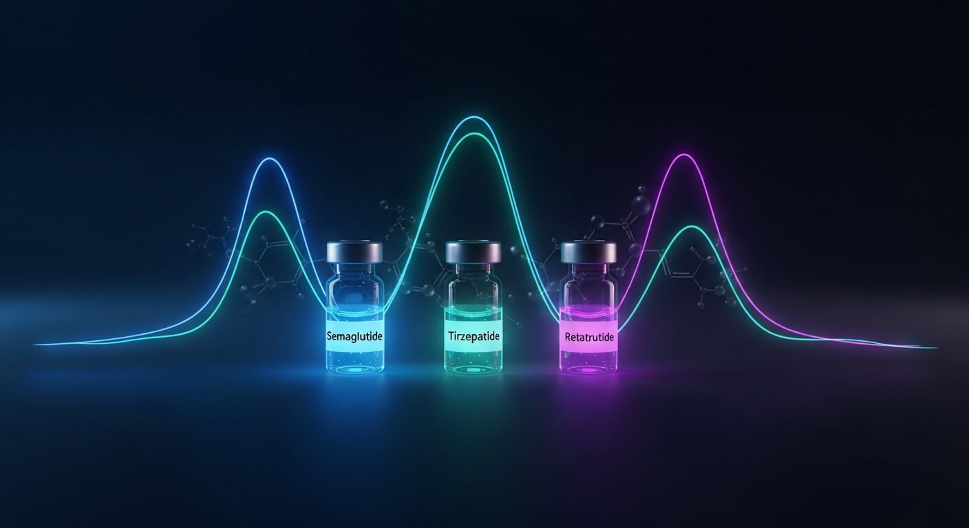
The three GLP-1 agonists at the top of the table (semaglutide, tirzepatide, retatrutide) have half-lives measured in days while most peptides are measured in minutes to hours. This is entirely due to engineering.

Native GLP-1 has a half-life of ~2 minutes — it’s rapidly degraded by DPP-4 enzymes. The research compounds achieve 5-7 day half-lives through two modifications: DPP-4-resistant amino acid substitutions (preventing enzymatic degradation) and fatty acid acylation that promotes albumin binding (preventing renal clearance).

They reach steady state in 4-5 weeks. If you’re measuring acute pharmacological effects, you need to account for this long wash-in period. CertaPeptides carries all three — see semaglutide, tirzepatide, and retatrutide.

Short-acting peptides: why minutes doesn’t mean ineffective

BPC-157’s ~2-minute IV half-life seems absurdly short. But half-life and efficacy aren’t the same thing. BPC-157’s reported effects have been linked to the NO system and to FAK-paxillin pathway activation (Sikiric et al., 2016, PMID: 27138887).

Similarly, Selank and Semax have half-lives under 3 minutes in their native forms, yet their effects on BDNF and NGF expression persist for hours. The peptide is the trigger, not the sustained effector.

Reconstituted solutions should be stored at 2-8°C and used within 28 days to maintain potency. Use our reconstitution calculator to work out concentrations, and check the peptide storage guide for detailed handling instructions.

How half-life affects storage stability

There’s a loose correlation between half-life and solution stability, but it’s not direct. Short half-life usually means the molecule is susceptible to enzymatic or chemical degradation — and those same vulnerabilities apply in the vial. Peptides with multiple basic residues (Arg, Lys) or free thiol groups (Cys) tend to degrade faster in solution.

General rules: lyophilized peptides at -20°C are stable for 6-24 months. Reconstituted peptides at 2-8°C last 21-28 days. Room temperature reconstituted peptides can lose significant activity within days. The shorter the half-life in vivo, the more critical proper storage becomes — because the same molecular vulnerabilities that cause rapid clearance also cause rapid degradation.

Frequently asked questions

What peptide has the longest half-life?

CJC-1295 with DAC at approximately 8 days, followed by semaglutide at ~7 days and cagrilintide at ~7 days. All three achieve long half-lives through albumin binding — DAC via covalent bonding, the others via fatty acid acylation. Without these modifications, native GLP-1 and native GHRH both have half-lives under 5 minutes.

What role does half-life play in experimental sampling design?

Half-life is one input into experimental sampling design but does not by itself determine it. BPC-157 (~2-minute half-life) is cleared rapidly; its reported effects are attributed to triggering downstream cascades rather than sustained receptor occupancy. The relationship between half-life and exposure depends on the mechanism of action.

Why is CJC-1295 DAC half-life so much longer than CJC-1295 without DAC?

The Drug Affinity Complex (DAC) covalently bonds to serum albumin after injection. Albumin has a ~19-day half-life in humans, so the peptide-albumin complex persists in circulation for days instead of minutes. Without DAC, CJC-1295 is a ~3 kDa peptide that’s rapidly cleared by the kidneys — below the ~60 kDa glomerular filtration threshold. With DAC, it becomes part of a ~70 kDa albumin complex that’s too large for renal clearance.

Peptide half-lives range from seconds (Selank, Semax) to 8 days (CJC-1295 DAC). GLP-1 agonists: semaglutide ~7 days, tirzepatide ~5 days, retatrutide ~6 days — all via albumin binding. GH secretagogues: ipamorelin ~2 hours, GHRP-2/6 ~15-25 minutes, MK-677 ~5 hours. Healing peptides: BPC-157 ~2 min IV (downstream effects persist), TB-500 est. 2-4 hours. Short half-life does not mean low activity in a model — mechanism of action matters as much as clearance.

Limitations of this reference

  • Many research peptides lack published human pharmacokinetic data. Values marked “Est.” are derived from animal studies or community reports and should be treated as approximations.
  • Half-life varies by route of administration, individual metabolism, and co-administered compounds. Values shown are typical for the most common research route.
  • This chart does not constitute dosing guidance. Research protocol design should reference primary literature for the specific compound and application.

References

  1. Lau J, et al. “Discovery of the Once-Weekly Glucagon-Like Peptide-1 (GLP-1) Analogue Semaglutide.” J Med Chem. 2015;58(18):7370-7380. PMID: 26308095
  2. Teichman SL, et al. “Prolonged stimulation of growth hormone (GH) and insulin-like growth factor I secretion by CJC-1295.” JCEM. 2006;91(3):799-805. PMID: 16352683
  3. Raun K, et al. “Ipamorelin, the first selective growth hormone secretagogue.” Eur J Endocrinol. 1998;139(5):552-561. PMID: 9849822
  4. Nass R, et al. “Effects of an oral ghrelin mimetic on body composition and clinical outcomes in healthy older adults.” Ann Intern Med. 2008;149(9):601-611. PMID: 18981485
  5. Sikiric P, et al. “Brain-gut Axis and Pentadecapeptide BPC 157: Theoretical and Practical Implications.” Curr Neuropharmacol. 2016;14(8):857-865. PMID: 27138887
  6. Dorr RT, et al. “Evaluation of melanotan-II, a superpotent cyclic melanotropic peptide in a pilot phase-I clinical study.” Life Sci. 1996;58(20):1777-1784. PMID: 8637402

All compounds discussed are for laboratory and educational research purposes only. Not for human consumption.

Posted on — Leave a comment

GLP-1 Peptide Pharmacokinetics: Why Molecular Modifications Shape Research Design

Introduction: why pharmacokinetics matter more than mechanism alone

When evaluating GLP-1 class peptides for research, most attention goes to their receptor binding profiles and downstream signaling. But a peptide’s pharmacokinetic (PK) profile, how long it circulates, how it resists degradation, and how it distributes through tissues, often determines research outcomes more than mechanism alone.

Native GLP-1 has a plasma half-life of approximately 2 minutes. Semaglutide lasts roughly 7 days. That difference is not a minor tweak, it represents a fundamental re-engineering of the molecule’s interaction with the body’s clearance systems. Understanding how these modifications work at the molecular level matters when designing rigorous research protocols.

This article examines the pharmacokinetic profiles of three GLP-1 class research peptides — semaglutide, tirzepatide, and retatrutide — with a focus on the molecular modifications that make each one unique.

All peptides discussed are for laboratory research purposes only and are not intended for human use.

The native GLP-1 problem

Glucagon-like peptide-1 (GLP-1) is a 30-amino acid incretin hormone secreted by intestinal L-cells in response to nutrient ingestion. It plays a central role in glucose homeostasis through multiple mechanisms: stimulating insulin secretion, suppressing glucagon release, slowing gastric emptying, and modulating appetite-related signaling in the central nervous system.

However, native GLP-1 faces three critical pharmacokinetic barriers that severely limit its research utility:

DPP-4 degradation

Dipeptidyl peptidase-4 (DPP-4) cleaves native GLP-1 at the alanine-2 position within minutes of secretion. This enzymatic degradation is the primary reason for the peptide’s approximately 2-minute plasma half-life. The cleavage product, GLP-1(9-36), has minimal receptor binding activity (Deacon et al., 1995).

Renal clearance

Even intact GLP-1 molecules that escape DPP-4 are rapidly cleared by the kidneys. The peptide’s small molecular weight (approximately 3,300 Da) falls well below the glomerular filtration threshold, allowing efficient renal elimination.

Short receptor occupancy

The combined effect of rapid degradation and clearance means native GLP-1 occupies its target receptor for only seconds to minutes, far too brief for sustained downstream signaling in research models requiring extended observation periods.

These limitations drove the development of modified GLP-1 analogs with engineered pharmacokinetic properties. Each of the three peptides discussed below solves these problems through different molecular strategies.

Semaglutide: C-18 fatty acid acylation

Molecular modifications

Semaglutide is a 31-amino acid GLP-1 receptor agonist with three key modifications that extend its half-life from 2 minutes to approximately 7 days, a 5,000-fold increase:

1. Aib8 substitution: The native alanine at position 8 is replaced with alpha-aminoisobutyric acid (Aib), which sterically blocks DPP-4 from accessing its cleavage site. This single substitution dramatically reduces enzymatic degradation (Lau et al., 2015).

2. C-18 fatty diacid chain: A C-18 octadecanedioic fatty acid is attached at lysine-26 via a mini-PEG linker. This modification is the primary driver of semaglutide’s extended half-life.

3. Arg34 substitution: Lysine at position 34 is replaced with arginine to prevent unwanted acylation at that site.

How albumin binding extends half-life

The C-18 fatty acid chain confers strong, reversible binding to serum albumin (approximately 99.9% bound). This albumin association provides three pharmacokinetic advantages:

  • Protection from DPP-4: The albumin-bound complex is too large and sterically hindered for DPP-4 to access the cleavage site
  • Reduced renal clearance: Albumin (66.5 kDa) is well above the glomerular filtration threshold, preventing renal elimination of the bound peptide
  • Depot effect: The equilibrium between bound and free semaglutide creates a slow-release depot, maintaining steady-state plasma concentrations over days

Semaglutide’s terminal half-life in humans is approximately 165 hours (approximately 7 days).

Tirzepatide: C-20 fatty diacid with dual agonism

Molecular modifications

Tirzepatide is a 39-amino acid peptide that represents a different engineering approach. Rather than modifying native GLP-1, it is based on the GIP (glucose-dependent insulinotropic polypeptide) sequence with engineered GLP-1 receptor cross-reactivity. Its key modifications include:

1. GIP-based backbone: The primary sequence derives from native GIP, providing full GIP receptor agonism. Strategic amino acid substitutions at specific positions enable cross-binding to GLP-1 receptors with approximately 5-fold lower affinity than native GLP-1 (Coskun et al., 2018).

2. C-20 eicosanedioic fatty diacid: A 20-carbon fatty diacid chain (two carbons longer than semaglutide’s C-18) is attached at lysine-20 via a linker. This longer acyl chain provides enhanced albumin binding affinity.

3. Aib2 substitution: Similar to semaglutide’s DPP-4 resistance strategy, the position-2 alanine is replaced with Aib to prevent enzymatic cleavage.

Pharmacokinetic profile

Tirzepatide achieves a terminal half-life of approximately 5 days (approximately 116 hours). While shorter than semaglutide’s 7-day half-life, tirzepatide’s dual receptor engagement means the net biological effect per injection may differ in research models. The dual GIP/GLP-1 activation produces complementary downstream signaling through both receptor pathways simultaneously (Willard et al., 2020).

The C-20 fatty diacid chain provides approximately 99.5% albumin binding. The slightly lower binding fraction compared to semaglutide (99.9%) results in a marginally higher free fraction, which may contribute to the different PK profile despite the longer acyl chain.

Retatrutide: triple agonism with acylated half-life extension

Molecular modifications

Retatrutide is the newest entrant in this class, engineered as a triple agonist targeting GIP, GLP-1, and glucagon receptors simultaneously. Its molecular architecture combines elements from both predecessors:

1. GIP-based backbone with triple receptor activity: Like tirzepatide, retatrutide uses a GIP-derived primary sequence but is further engineered to activate the glucagon receptor (GCGR) in addition to GIP-R and GLP-1R (Coskun et al., 2022).

2. Fatty acid acylation: An acyl chain provides albumin binding for half-life extension, following the same pharmacokinetic strategy as semaglutide and tirzepatide.

3. Optimized receptor balance: The relative potencies at each receptor are carefully tuned. Preclinical data suggests full GIP agonism, moderate GLP-1 agonism, and attenuated glucagon agonism, creating a specific signaling profile distinct from either single or dual agonists.

Pharmacokinetic profile

Retatrutide has a multi-day half-life comparable to semaglutide’s.

The addition of glucagon receptor agonism introduces a pharmacodynamic dimension not present in semaglutide or tirzepatide. Glucagon’s primary effects on hepatic glucose output and lipid oxidation create a metabolic signaling profile that differs qualitatively from incretin-only approaches.

Comparative pharmacokinetic table

Parameter Semaglutide Tirzepatide Retatrutide
Amino acids 31 39 39
Receptor targets GLP-1R only GIP-R + GLP-1R GIP-R + GLP-1R + GCGR
Primary sequence basis GLP-1 GIP GIP
Acyl chain C-18 fatty diacid C-20 fatty diacid Fatty acid (acylated)
DPP-4 resistance Aib8 substitution Aib2 substitution Engineered resistance
Half-life ~7 days (165 h) ~5 days (116 h) ~6-7 days (weekly dosing)
Albumin binding ~99.9% ~99.5% High (acylation-mediated)
Molecular weight ~4,114 Da ~4,810 Da ~4,500 Da (est.)
Phase of development Approved (research analog) Approved (research analog) Phase 3 clinical trials

How PK differences affect research protocol design

Steady-state accumulation

Peptides with longer half-lives accumulate to higher steady-state concentrations over repeated dosing. Semaglutide, with its 7-day half-life, takes roughly four to five half-lives to reach steady state. Tirzepatide, with a shorter 5-day half-life, reaches steady state slightly faster. Researchers designing multi-week studies must account for this accumulation phase when evaluating time-to-effect data.

Washout periods

The general pharmacological guideline is that 5 half-lives are required for near-complete elimination. For semaglutide, this means approximately 35 days of washout; for tirzepatide, approximately 25 days. Cross-over study designs must incorporate adequate washout periods to avoid carryover effects.

Reconstitution and handling

All three peptides are supplied as lyophilized powders requiring reconstitution before use. For solvent selection guidance, see our reconstitution guide.

Key takeaways

  • Native GLP-1 is pharmacokinetically impractical for research (2-minute half-life) — all three analogs solve this through fatty acid acylation and DPP-4-resistant modifications
  • Semaglutide uses a C-18 fatty diacid on a GLP-1 backbone for single-receptor, 7-day half-life
  • Tirzepatide uses a C-20 fatty diacid on a GIP backbone with engineered GLP-1R cross-reactivity for dual agonism, 5-day half-life
  • Retatrutide adds glucagon receptor agonism to the dual agonist formula, creating a triple receptor profile with weekly dosing
  • Albumin binding is the shared mechanism driving extended half-lives — the acyl chain length and structure determine binding affinity and free fraction
  • Pharmacokinetic modelling must account for accumulation kinetics and washout periods

Frequently asked questions

Q: Why does native GLP-1 have such a short half-life?

A: Native GLP-1 is rapidly degraded by the enzyme DPP-4, which cleaves the peptide at position 2 within minutes of secretion. Combined with rapid renal clearance due to its small molecular weight (approximately 3,300 Da), the resulting plasma half-life is only about 2 minutes. This is a normal physiological design, GLP-1 acts as a local paracrine signal, not a sustained circulating hormone.

Q: How does fatty acid acylation extend peptide half-life?

A: Attaching a fatty acid chain (C-18 or C-20) to the peptide enables reversible binding to serum albumin, a large protein (66.5 kDa) that circulates with a half-life of approximately 19 days. While bound to albumin, the peptide is protected from both DPP-4 degradation and renal filtration. The equilibrium between bound and free peptide creates a slow-release effect that maintains plasma concentrations over days.

Q: Why does tirzepatide have a shorter half-life than semaglutide despite having a longer acyl chain?

A: The relationship between acyl chain length and half-life is not strictly linear. Tirzepatide’s C-20 chain provides strong albumin binding, but its GIP-based backbone and different amino acid sequence may affect overall molecular stability, receptor-mediated internalization rate, and tissue distribution differently than semaglutide’s GLP-1-based structure. The net half-life reflects all these factors combined.

Q: What is the advantage of triple agonism over single or dual agonism?

A: Each additional receptor target introduces a distinct signaling pathway. GLP-1R drives incretin effects and appetite signaling. GIP-R adds complementary nutrient-sensing and adipose tissue signaling. GCGR (glucagon receptor) introduces hepatic glucose mobilization and lipid oxidation pathways. The hypothesis is that activating all three produces metabolic effects that are qualitatively different from, not simply additive to, single or dual agonist approaches.

Q: How should researchers account for accumulation when designing studies?

A: For weekly-dosed peptides with 5-7 day half-lives, steady state is reached after approximately 4-5 administrations. Studies measuring acute effects should distinguish between single-dose PK and steady-state PK data. For cross-over designs, a washout period of at least 5 half-lives (25-35 days) is recommended to avoid carryover confounds.

Q: Can these peptides be reconstituted with the same solvents?

A: Yes, all three are supplied as lyophilized powders. They are generally stable and tolerant of standard aqueous solvents. Store reconstituted solutions at 2-8°C and use within 28 days. See our reconstitution guide for detailed solvent recommendations.

References

  1. Deacon CF, Johnsen AH, Holst JJ. “Degradation of glucagon-like peptide-1 by human plasma in vitro yields an N-terminally truncated peptide that is a major endogenous metabolite in vivo.” The Journal of Clinical Endocrinology & Metabolism. 1995;80(3):952-957. DOI: 10.1210/jcem.80.3.7883856 | PMID: 7883856
  2. Lau J, Bloch P, Schaffer L, et al. “Discovery of the Once-Weekly Glucagon-Like Peptide-1 (GLP-1) Analogue Semaglutide.” Journal of Medicinal Chemistry. 2015;58(18):7370-7380. DOI: 10.1021/acs.jmedchem.5b00726 | PMID: 26308095
  3. Coskun T, Sloop KW, Loghin C, et al. “LY3298176, a novel dual GIP and GLP-1 receptor agonist for the treatment of type 2 diabetes mellitus: From discovery to clinical proof of concept.” Molecular Metabolism. 2018;18:3-14. DOI: 10.1016/j.molmet.2018.09.009 | PMID: 30473097
  4. Willard FS, Douros JD, Gabe MBN, et al. “Tirzepatide is an imbalanced and biased dual GIP and GLP-1 receptor agonist.” JCI Insight. 2020;5(17):e140532. DOI: 10.1172/jci.insight.140532 | PMID: 32730231
  5. Jastreboff AM, Kaplan LM, Frias JP, et al. “Triple-Hormone-Receptor Agonist Retatrutide for Obesity – A Phase 2 Trial.” New England Journal of Medicine. 2023;389(6):514-526. DOI: 10.1056/NEJMoa2301972 | PMID: 37366315
  6. Coskun T, Urva S, Roell WC, et al. “LY3437943, a novel triple glucagon, GIP, and GLP-1 receptor agonist for glycemic control and weight loss: From discovery to clinical proof of concept.” Cell Metabolism. 2022;34(9):1234-1247.e9. DOI: 10.1016/j.cmet.2022.07.013 | PMID: 35985340

Disclaimer

This article is for educational and research purposes only. The information provided does not constitute medical advice, dosing recommendations, or treatment guidance. Semaglutide, tirzepatide, and retatrutide are research peptides sold by CertaPeptides strictly for laboratory use and are not intended for human consumption. Always consult qualified professionals before beginning any research protocol.

Posted on — Leave a comment

GHK-Cu in 2026: Why Copper Peptide Research Interest Surged +1,016% Year-Over-Year

Disclaimer: This article is for educational and research purposes only. GHK-Cu peptides sold by CertaPeptides are intended strictly for in vitro research and laboratory use. This content does not constitute medical advice and should not be interpreted as an endorsement of human therapeutic use.

In a field dominated by GLP-1 compounds, one copper-binding tripeptide has quietly staged the most remarkable growth story of 2026. GHK-Cu — glycyl-L-histidyl-L-lysine copper(II) — saw its search interest surge +1,016% year-over-year, making it the single fastest-growing peptide in global search data according to The Peptide Effect’s 2026 State of peptides report.

To put that in perspective: GLP-1 peptides like semaglutide and tirzepatide still command roughly 60% of all peptide-related search traffic. But GHK-Cu’s growth trajectory is steeper than where those GLP-1 peptides were back in 2022, before they became household names. Researchers and institutions are paying attention — and the reasons why reveal where peptide Science is heading next.

The +1,016% surge — what’s driving GHK-Cu interest

Two converging forces explain why GHK-Cu research interest exploded in 2026.

The longevity research movement has reached mainstream velocity. Researchers and thought leaders like David Sinclair and Peter Attia have brought aging biology into public discourse, creating demand for compounds with documented roles in tissue repair and regeneration. GHK-Cu, first isolated from human plasma by Loren Pickart in 1973, fits squarely into this research paradigm as a naturally occurring peptide that declines with age (Pickart, 2008).

Published literature reached critical mass. With over five decades of peer-reviewed research, GHK-Cu has one of the deepest evidence bases of any bioactive peptide. The compound’s multi-target mechanism of action makes it particularly attractive for researchers exploring regenerative biology, wound healing models, and age-related tissue decline.

Understanding GHK-Cu — mechanism of action

GHK-Cu is a tripeptide with the amino acid sequence glycine-histidine-lysine, which forms a high-affinity complex with copper(II) ions. It occurs naturally in human plasma, saliva, and urine, with plasma concentrations declining significantly from approximately 200 ng/mL at age 20 to 80 ng/mL by age 60 (Pickart et al., 2015).

The peptide’s biological activity operates through several well-documented mechanisms:

  • Collagen synthesis stimulation: GHK-Cu has been shown to upregulate collagen types I, III, and V in fibroblast cultures, contributing to extracellular matrix remodeling. Early work by Maquart et al. (1988) demonstrated that GHK-Cu stimulated both collagen synthesis and the accumulation of several extracellular matrix components including glycosaminoglycans.
  • Angiogenesis promotion: Research indicates GHK-Cu promotes the formation of new blood vessels, a critical process in wound healing and tissue repair models (Pollard et al., 2005).
  • Anti-inflammatory activity: The peptide has been observed to modulate inflammatory markers, reducing oxidative damage indicators including lipid peroxidation, and suppressing pro-inflammatory cytokines such as TGF-beta and TNF-alpha in experimental models.
  • Antioxidant defense: GHK-Cu appears to support endogenous antioxidant systems, with studies suggesting it can modulate the expression of superoxide dismutase (SOD) and other protective enzymes.
  • Gene expression modulation: Broad gene expression studies have revealed that GHK-Cu may influence the activity of over 4,000 human genes, with a notable pattern of resetting gene expression toward a younger, healthier profile (Pickart et al., 2015).

The copper ion is not merely a structural component — it plays an active role in GHK-Cu’s biological activity. Copper is a cofactor for numerous enzymes involved in connective tissue formation, including lysyl oxidase, which is essential for collagen and elastin crosslinking.

Key research areas in 2026

The breadth of GHK-Cu research spans several active domains, each contributing to the compound’s surging interest.

Skin regeneration and anti-aging research

Dermatological research represents the most established application area for GHK-Cu. Studies have demonstrated the peptide’s ability to improve skin elasticity, firmness, and thickness in experimental models. Leyden et al. (2002) published findings showing that GHK-Cu facial cream improved skin laxity, clarity, and reduced fine lines in clinical observation studies. Researchers in 2026 are building on this foundation, investigating GHK-Cu’s role in photoaging models and UV damage repair pathways.

For researchers exploring synergistic peptide approaches, combinations with wound-healing peptides like BPC-157 and tissue-repair compounds like TB-500 have garnered interest.

Wound healing research

GHK-Cu’s wound healing properties have been documented since the 1980s. The peptide attracts immune cells and fibroblasts to wound sites, stimulates collagen deposition, and promotes angiogenesis — the trifecta of effective wound repair. Maquart et al. (1988) provided seminal evidence that GHK-Cu enhanced wound contraction and increased the synthesis of collagen, glycosaminoglycans, and decorin in rat wound models.

Current research extends these findings into chronic wound models, diabetic wound healing, and post-surgical recovery paradigms where impaired tissue repair mechanisms are a primary research challenge.

Hair follicle research

A growing body of research investigates GHK-Cu’s effects on hair follicle biology. The peptide’s ability to stimulate dermal papilla cells, promote angiogenesis around follicular structures, and modulate growth factors like VEGF and FGF has positioned it as a compound of interest in hair biology research. Studies suggest GHK-Cu may extend the anagen (growth) phase of the hair cycle and increase follicle size in experimental models.

Neuroprotection research

An emerging and rapidly growing research area examines GHK-Cu’s potential neuroprotective properties. Preclinical studies have explored the peptide’s antioxidant and anti-inflammatory effects in the context of neural tissue, with particular interest in its ability to modulate iron and copper homeostasis in the brain — metals implicated in neurodegenerative processes. While this research is still in early stages, the peptide’s ability to cross certain biological barriers and its documented gene expression effects have attracted NeuroScience laboratories to the field.

GHK-Cu vs other copper peptides

Not all copper peptides are equivalent, and researchers should understand the distinctions when designing experiments.

Compound Structure Copper Binding Research Profile
GHK-Cu Tripeptide (Gly-His-Lys) + Cu²⁺ High-affinity, specific 50+ years of published research; wound healing, collagen, gene expression
AHK-Cu Tripeptide (Ala-His-Lys) + Cu²⁺ Moderate affinity Limited literature; primarily cosmetic industry studies
Copper Gluconate Simple copper salt Non-specific Nutritional supplement; no peptide-mediated signaling
Copper Chloride Inorganic salt Non-specific Industrial/laboratory reagent; not bioactive peptide

GHK-Cu is distinguished by its specific copper-binding geometry and the extensive body of published research supporting its biological activity. The histidine residue provides the primary copper coordination site through its imidazole nitrogen, creating a stable yet bioavailable copper complex. This specificity is critical — generic copper supplementation does not replicate the targeted signaling effects observed with GHK-Cu in research models.

When sourcing GHK-Cu for research, the copper-peptide complex integrity is paramount. Improperly synthesized GHK-Cu may contain free copper ions, uncomplexed peptide, or degradation products that compromise experimental reproducibility.

Quality standards for GHK-Cu research

Reproducible research outcomes depend on peptide quality. For GHK-Cu specifically, several quality parameters deserve attention:

HPLC Purity Analysis: High-performance liquid chromatography remains the gold standard for peptide purity assessment. Research-grade GHK-Cu should demonstrate purity of 98% or higher by HPLC, ensuring minimal interference from synthesis byproducts, truncated sequences, or degradation products.

Mass Spectrometry Confirmation: Electrospray ionization mass spectrometry (ESI-MS) or MALDI-TOF confirms the molecular identity of the peptide, verifying the correct molecular weight of the GHK-Cu complex (approximately 403.9 Da for the copper-bound form). This step rules out structural analogs or misidentified compounds.

Copper Content Verification: Unlike most peptides, GHK-Cu requires verification of proper copper complexation. Inductively coupled plasma (ICP) analysis or atomic absorption spectroscopy can confirm stoichiometric copper binding and rule out free copper contamination.

Batch-Specific Certificates of Analysis: Every lot of GHK-Cu used in research should come with a batch-specific COA documenting purity, identity, and appearance. Generic or undated COAs are insufficient for publication-quality research.

CertaPeptides provides GHK-Cu in 50mg and 100mg research vials. Its product page shows the class-level specification on record, and we send selected lots to Janoshik for independent testing.

Frequently asked questions

What is GHK-Cu and why is it trending in 2026?

GHK-Cu (glycyl-L-histidyl-L-lysine copper) is a naturally occurring copper-binding tripeptide first identified in human plasma in 1973. Its search interest surged +1,016% year-over-year in 2026 according to The Peptide Effect report, driven by the growing longevity research movement and an expanding published literature base spanning wound healing, skin regeneration, and gene expression research.

How does GHK-Cu differ from generic copper supplements?

GHK-Cu is a specific peptide-copper complex with targeted biological signaling activity, unlike copper salts (gluconate, chloride) that simply provide copper ions. The tripeptide sequence creates a precise coordination geometry that mediates specific cellular responses including collagen synthesis stimulation, anti-inflammatory signaling, and gene expression modulation. These effects are not replicated by non-peptide copper sources.

What purity should researchers look for in GHK-Cu?

Research-grade GHK-Cu should be 98% or higher purity by HPLC analysis, with identity confirmed by mass spectrometry. Additionally, proper copper complexation should be verified, and each vial should include a batch-specific Certificate of Analysis. CertaPeptides’ GHK-Cu comes in 50mg and 100mg formats; its product page shows the class-level specification on record, and we send selected lots to Janoshik for independent testing.

What are the primary research applications for GHK-Cu?

The four most active research areas for GHK-Cu in 2026 are: (1) skin regeneration and anti-aging biology, (2) wound healing and tissue repair models, (3) hair follicle biology and growth cycle research, and (4) emerging neuroprotection studies. The peptide’s multi-target mechanism of action makes it relevant across these diverse research domains.

References

  1. Pickart, L. (2008). The human tri-peptide GHK and tissue remodeling. Journal of Biomaterials Science, Polymer Edition, 19(8), 969-988. DOI: 10.1163/156856208784909435 | PMID: 18644225
  2. Pickart L, Vasquez-Soltero JM, Margolina A. (2015). GHK peptide as a natural modulator of multiple cellular pathways in skin regeneration. BioMed Research International, 2015, Article 648108. DOI: 10.1155/2015/648108 | PMID: 26236730
  3. Maquart, F. X., Pickart, L., Laurent, M., Gillery, P., Monboisse, J. C., & Borel, J. P. (1988). Stimulation of collagen synthesis in fibroblast cultures by the tripeptide-copper complex glycyl-L-histidyl-L-lysine-Cu2+. FEBS Letters, 238(2), 343-346. DOI: 10.1016/0014-5793(88)80509-X | PMID: 3169264
  4. Pollard, J. D., Quan, S., Kang, T., & Koch, R. J. (2005). Effects of copper tripeptide on the growth and expression of growth factors by normal and irradiated fibroblasts. Archives of Facial Plastic Surgery, 7(1), 27-31. DOI: 10.1001/archfaci.7.1.27 | PMID: 15655171
  5. Leyden, J., Stephens, T., & Finkey, M. (2002). Skin care benefits of copper peptide containing facial cream. American Academy of Dermatology 60th Annual Meeting.

All peptides sold by CertaPeptides are intended for research purposes only. Not for human consumption. Purchasers must be qualified researchers or institutions. Always consult relevant regulations in your jurisdiction before conducting peptide research.

Posted on — Leave a comment

Cosmetic Peptides for Skin Research: GHK-Cu, Argireline, Matrixyl and Beyond

Introduction

The skin is the body’s largest organ — and among the most studied targets in peptide research. Over the past two decades, a distinct class of bioactive molecules known as cosmetic peptides has emerged as a major focus in dermatological Science. These short-chain amino acid sequences interact with receptors, enzymes, and signaling pathways in skin tissue with remarkable specificity, offering researchers tools to probe mechanisms of collagen synthesis, cellular senescence, neuromuscular signaling, and wound repair at the molecular level.

This guide examines the four major functional categories of cosmetic peptides studied in the research literature — signal peptides, carrier peptides, neurotransmitter-inhibiting peptides, and enzyme-inhibiting peptides — and provides a detailed look at three of the most extensively researched compounds: GHK-Cu, Argireline (Acetyl Hexapeptide-3), and Matrixyl (Palmitoyl Pentapeptide-4). All content here is strictly for educational and research purposes.

The four functional categories of cosmetic peptides

Cosmetic peptides are typically classified by their primary mechanism of action in skin tissue. Understanding these categories provides a framework for evaluating the mechanistic literature.

1. signal peptides

Signal peptides operate by binding to cell surface receptors and triggering downstream gene expression changes. In fibroblast cultures, several signal peptides have been shown to upregulate genes encoding collagen types I, III, and IV, as well as fibronectin and elastin. The Matrixyl family (palmitoyl oligopeptides) represents the most studied class of signal peptides in cosmetic research. These molecules mimic fragments of the extracellular matrix — particularly collagen breakdown products — which the body interprets as a wound repair signal, prompting fibroblasts to synthesize new structural proteins.

2. carrier peptides

Carrier peptides function primarily as chelating and delivery agents. The most prominent example is GHK-Cu (glycyl-L-histidyl-L-lysine:copper(II)), which forms a stable complex with copper ions and facilitates their delivery into skin tissue. Copper is a cofactor for lysyl oxidase, the enzyme responsible for cross-linking collagen and elastin fibers. Beyond copper delivery, GHK-Cu has been shown to act as a potent signal molecule in its own right, modulating a broad array of gene expression pathways.

3. neurotransmitter-inhibiting peptides

This category includes peptides that interfere with acetylcholine release at the neuromuscular junction, thereby reducing the intensity of repeated facial muscle contractions. Argireline (Acetyl Hexapeptide-3) and SNAP-8 (Acetyl Octapeptide-3) are the primary research compounds in this class. Their mechanism is analogous — though distinct in its molecular target — to botulinum toxin, and the scientific literature has explored both their efficacy and safety profile in controlled in vitro and in vivo settings.

4. enzyme-inhibiting peptides

Enzyme-inhibiting peptides work by blocking the activity of proteases and other enzymes that degrade extracellular matrix components. Matrix metalloproteinases (MMPs) — particularly MMP-1 (collagenase), MMP-2 (gelatinase A), and MMP-9 (gelatinase B) — are upregulated by UV exposure, oxidative stress, and inflammatory signaling. Research peptides in this category, such as soybean-derived Bowman-Birk inhibitor fragments and synthetic MMP inhibitors, have been studied for their capacity to preserve collagen networks in aged or UV-damaged tissue models.

GHK-Cu: the copper peptide research benchmark

GHK-Cu (glycyl-L-histidyl-L-lysine complexed with copper) is arguably the most thoroughly investigated cosmetic peptide in the scientific literature. First identified by Loren Pickart in 1973 during his research on liver cell regeneration, the tripeptide GHK was later found to have extraordinary skin-related biological activity when complexed with copper(II) ions.

Collagen synthesis and wound healing

A landmark study by Pickart and Margolina (2018) published in Biomolecules catalogued GHK-Cu’s influence on human gene expression, finding upregulation across 31 genes associated with collagen synthesis and downregulation in genes linked to inflammatory cascades. Fibroblast culture studies have repeatedly demonstrated that GHK-Cu stimulates the production of collagen I, collagen III, and glycosaminoglycans — the scaffold components of healthy dermis.

Arul et al. (2005) demonstrated in a rat wound model that topical GHK-Cu application significantly accelerated wound closure rates compared to controls, with histological analysis showing denser collagen fiber organization and improved neovascularization at wound sites. The authors attributed this to GHK-Cu’s role in recruiting fibroblasts and promoting TGF-β signaling at the injury site.

Antioxidant and anti-inflammatory activity

Research by Canapp et al. (2003) in the Veterinary Surgery journal demonstrated that GHK-Cu modulated inflammatory cytokine profiles in wound tissue, reducing TNF-α and IL-6 expression. At the same time, the peptide enhanced the activity of superoxide dismutase (SOD) — a key antioxidant enzyme — suggesting dual protective and reparative functions in stressed tissue environments.

Gene expression modulation

Perhaps the most striking finding in the GHK-Cu research corpus comes from gene array analysis. A 2012 study by Pickart, Vasquez-Soltero, and Margolina in Organogenesis found that GHK-Cu reset the gene expression profile of aging fibroblasts toward a more youthful state across 59 gene families. The peptide appeared to reverse many of the transcriptomic changes associated with cellular senescence, including upregulating genes in the ubiquitin proteasome system and downregulating genes associated with inflammation and cancer progression.

CertaPeptides supplies GHK-Cu for in vitro and research applications. Its product page shows the class-level specification on record, and selected lots have an independent third-party report.

Argireline and SNAP-8: neurotransmitter-inhibiting peptides

Mechanism of action

Argireline (INCI: Acetyl Hexapeptide-3 or Acetyl Hexapeptide-8) is a synthetic hexapeptide derived from the N-terminal domain of SNAP-25 (synaptosomal-associated protein 25), a component of the SNARE complex responsible for vesicle docking and neurotransmitter exocytosis at the neuromuscular junction. By competing with SNAP-25 for binding sites on the SNARE complex, Argireline research models suggest it can reduce acetylcholine release, thereby decreasing the amplitude of muscle fiber contractions associated with repetitive facial movement.

SNAP-8 (Acetyl Octapeptide-3) extends the Argireline peptide by two additional amino acids, and its developers proposed that this elongation improves binding affinity to the SNARE complex, potentially producing stronger modulation of vesicular release at lower concentrations.

Key research findings

A foundational study by Blanes-Mira et al. (2002) published in the International Journal of Cosmetic Science examined Argireline’s effects in both in vitro neuronal cell cultures and a clinical measurement study involving 10 volunteers. The in vitro data showed dose-dependent inhibition of acetylcholine release, while the clinical study using standardized skin measurement techniques found a measurable reduction in wrinkle depth over 30 days of topical application.

Wang et al. (2013) conducted a comparative assessment of Argireline versus SNAP-8 formulations in reconstructed human skin models, finding that SNAP-8 at 10% concentration produced approximately 30% greater reduction in contraction amplitudes compared to equivalent Argireline concentrations — though the authors noted that neither peptide approached the magnitude of botulinum toxin’s effect under equivalent research conditions.

Both peptides appear to operate through a reversible, competitive mechanism, distinguishing their research profile from irreversible protease-based inhibitors. This reversibility has made them subjects of continued interest in safety pharmacology research.

Penetration and delivery challenges

A consistent finding in the literature is that peptide size and charge significantly limit transdermal penetration. Research by Gorouhi and Maibach (2009) in Skin Pharmacology and Physiology reviewed the evidence base for topical peptides broadly and noted that unmodified peptides face considerable barrier function resistance from the stratum corneum. The acetylation of Argireline’s N-terminus improves lipophilicity somewhat, and current research directions include encapsulation in nanoparticle carriers, liposomes, and ethosomes to improve dermal delivery efficiency.

Matrixyl: signal peptide collagen research

Palmitoyl pentapeptide-4 (Matrixyl)

Matrixyl is the trade name for palmitoyl pentapeptide-4, a peptide comprising the sequence Lys-Thr-Thr-Lys-Ser (KTTKS) attached to a palmitoyl fatty acid chain via the lysine residue. The KTTKS sequence is derived from the pro-collagen I molecule — specifically from a domain that becomes exposed during collagen degradation. This places Matrixyl firmly in the signal peptide class: it functions as a matrikine, a fragment that signals to fibroblasts that collagen has been damaged and needs to be replaced.

Research on collagen I and III stimulation

Robinson et al. (2005) in the International Journal of Cosmetic Science conducted one of the most cited clinical studies on Matrixyl, enrolling 93 women in a double-blind, vehicle-controlled trial. The study measured wrinkle depth via optical profilometry and found statistically significant reductions at 12 weeks in the Matrixyl-treated group compared to vehicle. Ex vivo skin biopsy analysis from a parallel cohort demonstrated measurable increases in both procollagen I (27% above vehicle) and hyaluronic acid synthesis.

Katayama et al. (1993) earlier established the mechanistic basis for this class, demonstrating that the KTTKS sequence at nanomolar concentrations significantly upregulated collagen I, collagen III, and fibronectin synthesis in cultured human fibroblasts. The palmitoylation of Matrixyl increases the lipophilicity of the peptide and facilitates better interaction with cell membranes, which the research literature suggests improves delivery to dermal fibroblasts.

Matrixyl 3000: second-generation research

Matrixyl 3000 combines palmitoyl pentapeptide-4 with palmitoyl tetrapeptide-7 (Gly-Gln-Pro-Arg, a fragment of the Ig G molecule). Research by Lintner and Peschard (2000) demonstrated that the tetrapeptide component exerts anti-inflammatory activity by reducing IL-6 and TNF-α secretion in keratinocyte models, providing a complementary mechanism alongside the signal peptide’s collagen-stimulating effect. Studies comparing Matrixyl versus Matrixyl 3000 in fibroblast culture systems have generally reported enhanced collagen I and III gene expression with the combination compared to either component alone.

Comparative overview: key cosmetic peptides

Peptide Class Primary Mechanism Key Research Finding Molecular Weight
GHK-Cu Carrier / Signal Copper delivery; broad gene expression modulation Resets aging fibroblast transcriptome (Pickart 2012) 340.4 Da (free peptide)
Argireline Neurotransmitter-inhibiting Competes with SNAP-25 at SNARE complex Dose-dependent ACh release inhibition in vitro (Blanes-Mira 2002) 888.9 Da
SNAP-8 Neurotransmitter-inhibiting Extended SNARE complex competitor ~30% greater contraction reduction vs Argireline (Wang 2013) 1075.2 Da
Matrixyl (Pal-KTTKS) Signal (matrikine) Mimics pro-collagen fragment; stimulates ECM synthesis +27% procollagen I vs vehicle in clinical biopsy (Robinson 2005) 802.0 Da
Matrixyl 3000 Signal + Anti-inflammatory Pal-KTTKS + Pal-GQPR combination Enhanced collagen I/III vs single component (Lintner 2000) Combination
Leuphasyl Neurotransmitter-inhibiting Targets enkephalin receptors upstream of ACh release Synergistic effect with Argireline in combined models 655.8 Da

Market context: the cosmetic peptide research field

The global cosmetic peptide market has grown substantially over the past decade. According to market analysis reports from 2024, the cosmetic peptides sector was valued at approximately USD 1.2 billion and is projected to grow at a compound annual growth rate of 8–10% through 2030, driven by increased investment in active ingredient research, growing demand from the prestige skincare segment, and expanding regulatory interest in evidence-based cosmetic claims.

This growth has been paralleled by a surge in academic and industry research. PubMed indexed approximately 340 cosmetic peptide-related publications in 2010; by 2023, that figure had grown to over 1,200 annual publications. The majority focus on three themes: mechanism elucidation in cell culture models, transdermal delivery optimization, and head-to-head efficacy comparisons using standardized outcome measures such as optical profilometry, ultrasound dermometry, and confocal microscopy.

Key research trends as of 2025-2026 include:

  • Combination approaches: Studies increasingly examine synergistic peptide combinations rather than single-compound models, reflecting the multi-pathway Nature of skin aging.
  • Delivery innovation: Nanostructured lipid carriers, peptide-loaded microneedle patches, and exosome-based delivery systems are being actively investigated to overcome the permeability barrier.
  • Biomarker standardization: The field is moving toward consensus biomarker panels (procollagen I, MMP-1 activity, hyaluronic acid synthase expression) to enable cross-study comparisons.
  • Genomic approaches: GHK-Cu’s demonstrated ability to modulate hundreds of genes simultaneously has opened interest in “epigenetic peptides” — compounds that may influence aging trajectories at the transcriptional level.

Research protocols and handling considerations

Researchers working with cosmetic peptides in laboratory settings should be aware of several practical considerations.

Reconstitution

Most lyophilized cosmetic peptides reconstitute readily in aqueous buffers at physiological pH (7.2–7.4). GHK-Cu, due to its copper complex, should be reconstituted in water or phosphate-buffered saline; avoid chelating buffers (e.g., EDTA-containing solutions) which can strip the copper ion. Argireline and Matrixyl are generally stable in aqueous solution at neutral pH and can be dissolved at 1–10 mg/mL stock concentrations for dilution into cell culture media.

Storage

Lyophilized cosmetic peptides should be stored desiccated at -20°C in single-use aliquots to prevent freeze-thaw degradation. GHK-Cu specifically should be protected from light exposure, as copper-catalyzed oxidation can occur under UV illumination. Once reconstituted, working solutions are typically stable for 24–72 hours at 4°C; for longer storage, prepare fresh aliquots from lyophilized stock.

In vitro model systems

The predominant research models for cosmetic peptides include: primary human dermal fibroblast (HDF) cultures for collagen synthesis assays; reconstructed human epidermis (RHE) models such as EpiDerm for barrier function studies; and ex vivo human skin explants for more physiologically relevant penetration and activity data. Researchers should note that results from 2D cell culture may not directly translate to 3D tissue models, and interpretation across model systems requires careful consideration of peptide concentration, exposure time, and the specific endpoints measured.

Internal research resources

For researchers exploring this area, the following CertaPeptides resources may be relevant:

Frequently asked questions

What are cosmetic peptides used for in research?

In research contexts, cosmetic peptides are used to investigate mechanisms of skin aging, collagen synthesis regulation, neuromuscular signaling in skin tissue, and extracellular matrix remodeling. They serve as molecular tools to interrogate specific biological pathways in fibroblast cultures, reconstructed skin models, and ex vivo tissue systems. This content is for educational and research purposes only.

How does GHK-Cu differ from other cosmetic peptides?

GHK-Cu is classified as a carrier peptide due to its ability to chelate and transport copper(II) ions, but research demonstrates it also functions as a potent signal peptide with broad gene expression effects. Unlike signal peptides such as Matrixyl that mimic specific ECM fragments, GHK-Cu modulates hundreds of gene families simultaneously, including pathways related to inflammation, antioxidant defense, DNA repair, and collagen synthesis.

What is the research significance of argireline’s SNARE mechanism?

Argireline’s proposed mechanism — competitive inhibition of SNAP-25 at the SNARE complex — is significant because it targets the same vesicular release machinery as botulinum toxin but through a different binding site and with a reversible, competitive mechanism. This makes Argireline a useful research tool for studying neuromuscular junction pharmacology in skin-relevant models and for exploring reversible approaches to neurotransmitter modulation.

Are cosmetic peptides stable in cell culture conditions?

Stability varies by peptide. In general, short peptides below 500 Da may be subject to rapid enzymatic degradation by serum proteases in cell culture media containing fetal bovine serum. Researchers typically use serum-free or reduced-serum conditions for short-duration mechanistic studies, or verify stability via HPLC analysis of conditioned media. Longer or modified peptides (e.g., acylated peptides like Matrixyl) tend to show improved stability due to protease resistance conferred by the fatty acid modification.

What is the difference between matrixyl and matrixyl 3000?

Matrixyl refers to palmitoyl pentapeptide-4 (Pal-KTTKS) alone, a matrikine signal peptide that stimulates collagen I and III synthesis by mimicking a pro-collagen degradation fragment. Matrixyl 3000 is a combination of palmitoyl pentapeptide-4 with palmitoyl tetrapeptide-7 (Pal-GQPR), which adds an anti-inflammatory component targeting IL-6 and TNF-alpha pathways. Research in fibroblast and keratinocyte models suggests the combination produces synergistic effects on extracellular matrix gene expression.

How do researchers measure the efficacy of cosmetic peptides?

Standard research outcome measures include: quantitative RT-PCR for gene expression (COL1A1, COL3A1, MMP-1, HAS2); ELISA or Western blot for protein quantification of procollagen I, collagen III, and fibronectin; Sircol collagen assay for total soluble collagen in conditioned media; optical profilometry and confocal microscopy for morphological assessment; and standardized wrinkle depth scoring (Antera 3D, VISIA, or similar) in clinical study designs. Each method has distinct sensitivity thresholds and interpretation requirements.

What concentration ranges are typically used in cosmetic peptide research?

Concentrations in the literature span a wide range depending on the experimental model. In fibroblast culture studies, GHK-Cu is commonly studied at 1 nM to 1 µM; Argireline typically at 100 µM to 1 mM for in vitro neurotransmitter release assays; and Matrixyl at 1–100 µM for collagen synthesis models. These concentrations are established for in vitro research purposes only and do not represent any recommendations for other applications.

How large is the cosmetic peptide research market?

Market analyses from 2024 estimate the global cosmetic peptides market at approximately USD 1.2 billion, with projected growth to USD 2.0–2.5 billion by 2030 at a CAGR of 8–10%. Growth is driven by increasing R&D investment in active cosmetic ingredients, rising consumer demand for evidence-based formulations, and the expansion of the prestige skincare segment globally. Academic publication rates on cosmetic peptides have more than tripled between 2010 and 2023.

Cosmetic peptides — classified as signal, carrier, neurotransmitter-inhibiting, or enzyme-inhibiting compounds — are increasingly important molecular tools in dermatological research. Of the compounds reviewed here, GHK-Cu has the deepest published evidence base, with documented effects spanning collagen synthesis stimulation, antioxidant gene regulation, and broad transcriptomic remodeling in fibroblast cultures. Argireline and SNAP-8 offer a reversible, competitive mechanism targeting the SNARE complex — distinct from botulinum toxin — making them useful research tools for investigating neuromuscular junction pharmacology. Matrixyl and Matrixyl 3000 represent the matrikine signal peptide class, with published clinical and in vitro data supporting their role in collagen I and III synthesis. As delivery Science advances and combination study designs become more prevalent, the cosmetic peptide research field continues to expand beyond its origins in single-compound efficacy assessment.

References

  1. Pickart L, et al. (2015). GHK peptide as a natural modulator of multiple cellular pathways in skin regeneration. BioMed Research International, 2015, 648108. PMID: 26236730.
  2. Blanes-Mira C, et al. (2002). A synthetic hexapeptide (Argireline) with antiwrinkle activity. International Journal of Cosmetic Science, 24(5), 303-310. PMID: 18498523.
  3. Robinson LR, et al. (2005). Topical palmitoyl pentapeptide provides improvement in photoaged human facial skin. International Journal of Cosmetic Science, 27(3), 155-160. PMID: 18492182.

Disclaimer: This article is for educational and research purposes only. The information provided does not constitute medical advice, and the compounds discussed are intended solely for laboratory and scientific research use. Always consult qualified professionals before beginning any research protocol. CertaPeptides products are not for human or veterinary use.

Posted on — Leave a comment

Bioregulator Peptides: The Complete Guide to Khavinson Peptides and Organ-Specific Research

What are bioregulator peptides?

Bioregulator peptides represent a distinct class of signaling molecules — ultra-short chains of just two to four amino acids — that research suggests play a fundamental role in regulating gene expression at the cellular level. Unlike larger peptides that act primarily through receptor binding on the cell surface, bioregulators are proposed to work intracellularly, interacting directly with chromatin and influencing which genes are transcribed in a given tissue.

The concept of peptide bioregulators emerged from decades of research into the molecular mechanisms of aging, tissue homeostasis, and organ-specific gene regulation. What distinguishes them from conventional peptides is their extraordinary target specificity: each bioregulator is hypothesized to exert preferential effects on a particular organ or tissue type, acting as what researchers describe as an “organ-specific” signaling agent.

This article provides an educational overview of the science, history, and current research context surrounding bioregulator peptides — for research and informational purposes only. Nothing in this article constitutes medical advice.

The history: 40+ years of Khavinson research

The systematic study of peptide bioregulators was pioneered by Professor Vladimir Khavinson, a Russian gerontologist and biochemist who began his work in the late 1960s at the St. Petersburg Institute of Bioregulation and Gerontology (part of the Russian Academy of Medical Sciences). Khavinson and his colleagues spent more than four decades investigating the biological activity of short peptide fractions extracted from animal organs and tissues.

The original research program was partly motivated by Soviet military and space interests: scientists sought methods to protect personnel from the accelerated aging effects of extreme stress, radiation, and demanding operational environments. Funding from these programs allowed for unusually large, long-duration clinical research programs that produced a substantial body of published data.

The early methodology involved extracting peptide fractions from calf thymus, pineal gland, liver, and other organs, then testing these extracts in cell culture, animal models, and eventually human clinical studies. Over time, the research evolved from crude tissue extracts toward the identification and synthesis of specific short peptide sequences — some as small as two amino acids — that appeared to account for the majority of the observed biological activity.

Khavinson has authored or co-authored over 800 peer-reviewed publications. His work spans from molecular biology and chromatin structure to longitudinal human studies tracking cohorts for 10 to 15 years. The St. Petersburg Institute remains the primary center for this field of research globally, though interest has expanded internationally, particularly in the EU and North America, over the past decade.

Proposed mechanism of action: gene expression regulation

The mechanism proposed for peptide bioregulators is distinct from most peptide research and is a key area of ongoing scientific investigation. Rather than binding to membrane receptors, bioregulators are proposed to penetrate cell membranes and enter the nucleus, where they interact with histones and specific DNA sequences.

Chromatin interaction model

Research by Khavinson et al. suggests that short peptides with specific amino acid sequences can bind to the major groove of DNA through complementary electrostatic interactions. The hypothesis is that each dipeptide or tripeptide sequence is “complementary” to a specific DNA promoter region — a concept Khavinson’s team has termed the “peptide-DNA complementarity” model.

According to this model, when a bioregulator binds to a promoter or enhancer region, it facilitates binding of transcription factors and RNA polymerase, effectively upregulating gene expression for proteins associated with cellular repair, differentiation, and maintenance. This mechanism, if validated at scale, would explain why the same dipeptide might produce different effects in different cell types — the accessible chromatin architecture varies by tissue, so the same molecule would activate different genes depending on its cellular context.

Epigenetic modulation

A parallel line of research focuses on the potential of bioregulators to influence epigenetic marks — specifically DNA methylation patterns and histone acetylation status. Studies indicate that Aging cells accumulate abnormal methylation patterns that suppress the expression of maintenance and repair genes. Research data suggests that certain short peptides may help restore more youthful methylation patterns in aged cell cultures, though the precise molecular mechanism remains under active investigation.

A 2014 study published in Advances in Gerontology (Khavinson et al.) reported that Epithalon — a tetrapeptide — caused measurable changes in telomerase activity and reduced methylation of specific gene promoters in cultured human cells. These findings have been cited as evidence for the epigenetic mechanism hypothesis, though replication in independent laboratories remains limited.

Key bioregulator peptides: research overview

The following sections summarize the primary bioregulators that have been subjects of published research. All data cited is from peer-reviewed sources and is presented for educational purposes.

Epitalon / epithalamin (pineal gland)

Epitalon (Ala-Glu-Asp-Gly, a tetrapeptide) is a synthetic version of Epithalamin, the naturally-occurring polypeptide fraction originally isolated from bovine pineal gland. It is the most extensively researched peptide bioregulator, with publications spanning from molecular cell biology to clinical longitudinal trials.

Research suggests Epitalon may influence melatonin synthesis through pineal gland signaling pathways. A study by Anisimov et al. (2003) published in Annals of the New York Academy of Sciences reported that mice administered Epitalon demonstrated a statistically significant 13% extension of mean lifespan compared to controls, along with reduced tumor incidence. The study included 210 animals across three cohorts and is frequently cited as one of the foundational longevity studies in this field.

A separate clinical study by Khavinson and Morozov (2003) tracked 266 elderly subjects over three years, with a subgroup receiving Epithalamin supplementation. Data indicated improvements in immune parameters (T-lymphocyte counts, NK cell activity), as well as cardiovascular markers. The study was published in Neuro Endocrinology Letters.

CertaPeptides offers Epitalon 50mg for research purposes. See also our guide: Epitalon and Telomere Research.

Thymalin / thymogen (thymus and immune system)

Thymalin is a polypeptide complex derived from calf thymus tissue, while Thymogen is a synthetic dipeptide (Glu-Trp) identified as one of the active components of Thymalin. Both have been studied primarily in the context of immune system regulation, particularly age-related immune decline (immunosenescence).

The data indicated a 2.0 to 2.4-fold reduction in mortality compared to untreated controls over the study period. Immune biomarkers measured included CD4/CD8 ratios, NK cell counts, and immunoglobulin levels. While methodological limitations of the study design limit generalizability, the long follow-up period is notable.

Research on Thymogen specifically has focused on its ability to stimulate differentiation of T-lymphocyte precursors. In vitro studies suggest the dipeptide enhances expression of CD3 and other T-cell markers, consistent with a role in supporting thymic output — a function known to decline substantially with age.

Pinealon (brain and CNS)

Pinealon is a tripeptide (Glu-Asp-Arg) studied primarily for its proposed neuroprotective and cognitive effects. Unlike Epitalon — which targets the pineal gland as an organ — Pinealon has been investigated for direct effects on neuronal cells themselves.

Khavinson et al. (2011) published a study in Rejuvenation Research examining Pinealon’s effects on cultured neuronal cells under conditions of oxidative stress. Data showed reduced free-radical levels, improved cell viability, and enhanced proliferative activity in peptide-treated cultures compared to controls. The authors proposed that Pinealon’s protective effects are consistent with the chromatin-interaction and gene-expression mechanism described above for the broader bioregulator peptide class.

A 2012 study (Grigoriev et al., published in Advances in Gerontology) examined Pinealon’s effects in a rat model of cerebral ischemia-reperfusion injury. Animals treated with Pinealon exhibited smaller infarct volumes and better performance on maze-based cognitive tests at 30 days post-injury compared to controls.

Vilon (immune regulation)

Vilon is a dipeptide (Lys-Glu) with a primary research focus on immune modulation, particularly in the context of aging and autoimmune conditions. Research suggests it may influence the balance between T-helper cell subpopulations (Th1/Th2), with potential relevance to inflammation regulation.

A study by Khavinson et al. (2002, Bulletin of Experimental Biology and Medicine) investigated Vilon in aged rats and reported normalization of cytokine profiles — specifically reduced IL-6 and TNF-alpha expression — in treated animals compared to age-matched controls. The authors interpreted this as evidence of an anti-inflammatory regulatory effect at the transcriptional level, consistent with the broader bioregulator mechanism hypothesis.

Livagen (liver and chromatin)

Livagen (Lys-Glu-Asp-Ala) is a tetrapeptide studied in the context of liver function and, notably, as a research tool for investigating chromatin remodeling. Its published research record is somewhat different from other bioregulators: Livagen has been used extensively in cell biology experiments studying how short peptides interact with nucleosomes and modify gene expression in hepatic tissue.

Khavinson et al. (2014, Frontiers in Bioscience) used Livagen in a mechanistic study of peptide-chromatin interaction, demonstrating via chromatin immunoprecipitation (ChIP) assays that the tetrapeptide enhanced acetylation of histone H3 at promoter regions of specific liver genes. This study is significant because it provides relatively direct molecular evidence — rather than just outcomes data — for the proposed epigenetic mechanism of bioregulators.

Testagen (testes and reproductive research)

Testagen (Lys-Asp-Glu-Glu) is a tetrapeptide proposed to act as an organ-specific bioregulator for testicular tissue. Research in this area has focused on age-related decline in testosterone production and spermatogenesis.

Animal studies (Khavinson et al., Advances in Gerontology, 2010) reported that aged male rats administered Testagen showed improvements in both Leydig cell function (as measured by testosterone output ex vivo) and sperm morphology parameters. The authors noted upregulation of StAR (steroidogenic acute regulatory protein) gene expression in treated testicular tissue, which they cited as consistent with the gene-expression regulatory mechanism.

Chonluten (bronchial and lung tissue)

Chonluten is a tripeptide (Gly-Glu-Pro) investigated primarily for potential effects on bronchial epithelial cell function. Research has focused on its influence on mucus secretion, ciliary activity, and epithelial cell regeneration after injury.

A study by Khavinson et al. (2006, Bulletin of Experimental Biology and Medicine) examined Chonluten in an in vitro model of bronchial epithelial cells exposed to oxidative stress. Peptide-treated cells showed significantly higher viability at 24 and 48 hours post-insult, along with enhanced expression of surfactant protein genes. The authors proposed Chonluten as a candidate for lung tissue regeneration research, though no in vivo data was presented in that particular publication.

Bioregulator comparison table

Bioregulator Sequence Length Target Organ/System Primary Research Area Key Study Reference
Epitalon Ala-Glu-Asp-Gly 4 AA Pineal Gland Longevity, telomere length, melatonin Anisimov et al. (2003)
Epithalamin Polypeptide complex ~30-40 AA Pineal Gland Immune function, cardiovascular markers Khavinson & Morozov (2003)
Thymalin Polypeptide complex Multiple Thymus / Immune T-cell differentiation, mortality Khavinson et al. (2002)
Thymogen Glu-Trp 2 AA Thymus / Immune T-lymphocyte differentiation Various, 1990s-2010s
Pinealon Glu-Asp-Arg 3 AA Brain / CNS Neuroprotection, cognitive function Grigoriev et al. (2012)
Vilon Lys-Glu 2 AA Immune System Cytokine modulation, inflammation Khavinson et al. (2002)
Livagen Lys-Glu-Asp-Ala 4 AA Liver Chromatin remodeling, hepatic gene expression Khavinson et al. (2014)
Testagen Lys-Asp-Glu-Glu 4 AA Testes / Reproductive Testosterone production, spermatogenesis Khavinson et al. (2010)
Chonluten Gly-Glu-Pro 3 AA Lungs / Bronchial Epithelial cell regeneration, surfactant Khavinson et al. (2006)

Research strengths and limitations

Any serious evaluation of bioregulator peptide research must acknowledge both its strengths and its limitations. Understanding these factors is essential for researchers and professionals reviewing this body of work.

Strengths

  • Volume and duration: The bioregulator research program is one of the most sustained peptide research efforts in history, spanning over 40 years with hundreds of publications in peer-reviewed journals.
  • Mechanistic specificity: The chromatin-interaction and epigenetic hypotheses are mechanistically coherent and testable, and some molecular evidence (e.g., ChIP assays from the Livagen studies) supports the framework.
  • Organ specificity hypothesis: The organ-targeting concept is scientifically interesting and, if validated, would represent a significant advance in targeted peptide research.
  • Longitudinal human data: Unlike most peptide research which relies exclusively on animal models, some bioregulator studies tracked human subjects for 10-15 years — a rarity in this field.

Limitations and considerations

  • Replication gap: The vast majority of published research originates from a single research group (Khavinson et al. at the St. Petersburg Institute). Independent replication in Western laboratories is sparse, limiting consensus validation.
  • Oral bioavailability questions: Short peptides administered orally face digestion in the gastrointestinal tract. Published studies have used various routes — subcutaneous, intranasal, and oral — with inconsistent bioavailability data across routes.
  • Mechanistic complexity: The peptide-DNA complementarity model, while internally coherent, has not yet been definitively demonstrated through high-resolution structural biology methods (e.g., X-ray crystallography of peptide-DNA complexes).
  • Study design variability: Earlier studies from the Soviet era were conducted under different methodological standards than contemporary randomized controlled trial (RCT) protocols, making cross-study comparison difficult.
  • Regulatory status: Bioregulator peptides are not approved therapeutic agents in the EU, US, or most jurisdictions. They are available strictly as research compounds.

Bioregulators vs. other peptide classes

Understanding how bioregulators differ from more widely studied peptide classes helps contextualize the research:

Bioregulators vs. growth hormone secretagogues (e.g., Ipamorelin, CJC-1295)

Growth hormone secretagogues work primarily through receptor-mediated signaling at the pituitary and hypothalamus, stimulating GH release. Their mechanism is well-characterized at the receptor level. Bioregulators, by contrast, are proposed to act downstream at the gene expression level and are not primarily growth hormone-related. The two classes are mechanistically distinct research tools.

See our related guides: Ipamorelin Research and CJC-1295 DAC: Complete Research Guide.

Bioregulators vs. tissue repair peptides (e.g., BPC-157, TB-500)

BPC-157 and TB-500 operate through well-documented receptor-mediated and cytoskeletal mechanisms respectively — BPC-157 via VEGFR2 and FAK/paxillin pathways, TB-500 through actin-sequestration and MRTF-A signaling. These are larger peptides (15 and 44 amino acids respectively) with more conventional pharmacological profiles. Bioregulators are fundamentally shorter and proposed to act by a different primary mechanism. Researchers studying tissue regeneration may be interested in both classes but should not conflate their mechanisms.

Related reading: BPC-157 vs. TB-500: Comprehensive Comparison Guide.

Bioregulators vs. anti-aging peptides (e.g., GHK-Cu)

GHK-Cu (copper tripeptide) is classified by some researchers as a “bioregulator-adjacent” compound due to its short length and gene-expression effects, but it operates through different pathways — primarily copper chelation, collagen synthesis signaling, and antioxidant gene upregulation via Nrf2. GHK-Cu’s mechanism is more extensively characterized in Western literature than classical Khavinson bioregulators. Both classes represent active areas of short-peptide gene-regulation research.

Practical research considerations

Storage and handling

Short peptides in general are chemically stable relative to larger peptides, owing to their smaller molecular size and reduced susceptibility to tertiary structure disruption. Research-grade bioregulator peptides are typically supplied in lyophilized (freeze-dried) form and should be stored according to the following general guidelines:

  • Lyophilized powder: Store at -20°C in a sealed, light-protected container for optimal long-term stability.
  • Reconstituted solution: Use bacteriostatic water for reconstitution; store at 4°C and use within 28-30 days.
  • Avoid repeated freeze-thaw cycles of reconstituted peptide; prepare aliquots if extended storage is needed.
  • Protect from light — particularly UV — as aromatic amino acid residues (e.g., Trp in Thymogen) can undergo photo-oxidation.

For a comprehensive protocol reference, see: Complete Peptide Storage Guide: Temperature Chart.

Reconstitution protocol

Reconstitution of lyophilized bioregulator peptides follows standard research peptide protocols. The short chain length of most bioregulators (2-4 amino acids) generally makes them highly water-soluble, simplifying reconstitution compared to longer, hydrophobic peptides. Researchers typically use bacteriostatic water at a ratio appropriate for the desired concentration, injecting the solvent slowly against the inner wall of the vial rather than directly onto the lyophilized cake to minimize mechanical degradation.

Frequently asked questions

What is the difference between epithalamin and Epitalon?

Epithalamin is the naturally occurring polypeptide complex extracted from bovine pineal gland tissue. It contains multiple peptide sequences of varying length. Epitalon is the synthetic tetrapeptide (Ala-Glu-Asp-Gly) identified by Khavinson’s team as the primary active sequence within Epithalamin. Epitalon is easier to synthesize with high purity and reproducibility, which is why most contemporary research uses the synthetic version. For research purposes, Epitalon is the more precisely defined compound.

Are bioregulator peptides the same as nootropic peptides like Semax or Selank?

No — they are distinct classes. Nootropic peptides like Semax (ACTH fragment) and Selank (tuftsin analogue) were developed through a different research lineage (primarily at the Russian Institute of Molecular Genetics) and work primarily through receptor-mediated mechanisms involving BDNF, serotonin, and dopamine pathways. Khavinson bioregulators are distinct in their proposed mechanism (chromatin/gene expression) and in having organ-specific rather than CNS-primary target profiles. Pinealon — a bioregulator — does have CNS research interest, but through a different proposed mechanism than Semax or Selank.

How does “organ-specificity” work for such small molecules?

This is one of the central scientific questions in bioregulator research. The proposed explanation is that organ-specificity emerges from the tissue-specific chromatin architecture rather than from selective biodistribution of the peptide itself. Different cell types have different patterns of open and closed chromatin — different gene promoters are accessible in a hepatocyte versus a thymic epithelial cell. The hypothesis is that a given bioregulator sequence is complementary to promoter regions of genes that happen to be accessible (open chromatin) primarily in its target organ, producing apparently organ-specific effects from what may be systemically distributed molecules. This model is elegant but remains incompletely validated in the literature.

Where is the research published?

Bioregulator research has been published across a range of journals including Bulletin of Experimental Biology and Medicine, Advances in Gerontology, Neuro Endocrinology Letters, Annals of the New York Academy of Sciences, and Frontiers in Bioscience. PubMed indexes many of these publications under search terms including “Khavinson”, “peptide bioregulators”, and the individual peptide names. The research originates primarily from Russian institutions, which is relevant context for evaluating its translation to Western regulatory frameworks.

Are bioregulator peptides orally bioavailable?

Bioavailability via the oral route is a significant research question. Published studies have used various administration routes. Some researchers propose that the ultra-short length (2-4 amino acids) of bioregulators may confer greater resistance to gastrointestinal peptidase digestion compared to longer peptides, though comprehensive pharmacokinetic studies with validated oral bioavailability data are limited. Subcutaneous and intranasal routes have been used in several published protocols. As with all research compounds, route of administration and dosing are matters for the research protocol design team to determine based on the specific experimental objectives.

What is the regulatory status of bioregulator peptides?

In the European Union, bioregulator peptides are classified as research compounds and are not approved as medicinal products. Some are registered as dietary supplements in Russia, where the research originated — Epithalamin and Thymalin, for example, have been registered pharmaceutical products in Russia since the 1980s. In the EU and US, they are available for research purposes only and are not approved for clinical use or human therapeutic application. Researchers should review the regulations applicable in their specific jurisdiction before initiating any research program involving these compounds.

How do bioregulators relate to the hallmarks of aging framework?

The hallmarks of aging framework (López-Otín et al., cell, 2013; updated 2023) identifies epigenetic alterations, loss of proteostasis, and cellular senescence as primary drivers of the aging phenotype. The proposed mechanism of bioregulators — restoring more youthful patterns of gene expression through chromatin interaction — maps conceptually onto the “epigenetic alterations” hallmark. Epitalon’s research record, which includes telomerase activation data, intersects with the “telomere attrition” hallmark. Whether bioregulators measurably address these hallmarks in vivo at physiologically relevant concentrations is an active area of research that has not yet been resolved in the Western scientific literature.

What does CertaPeptides offer in the bioregulator category?

CertaPeptides currently offers Epitalon 50mg for research purposes. Our catalogue continues to expand as we source additional research-grade compounds meeting our quality standards. All products are supplied for research purposes only; product pages show a class-level specification where one is on record, and we send selected lots to Janoshik for independent testing. View available products at Anti-Aging Research and Bioregulators.

Bioregulator peptides are ultra-short (2–4 amino acid) chains proposed to regulate gene expression through direct chromatin interaction rather than receptor-mediated signaling — a mechanism developed over 40+ years by Vladimir Khavinson and the St. Petersburg Institute, with hundreds of peer-reviewed publications. Each bioregulator is hypothesized to act on a specific organ: Epitalon/Epithalamin on the pineal gland, Thymalin/Thymogen on the thymus, Pinealon on the brain, Vilon on immune function, Livagen on the liver, Testagen on the testes, and Chonluten on the lung. Epitalon has the strongest evidence base, including telomerase activation findings and longitudinal human data. Key limitations to keep in mind: most published research originates from a single institution, oral bioavailability data is limited, and high-resolution structural confirmation of the peptide-DNA binding mechanism remains unpublished. All bioregulator peptides are research compounds only — not approved therapeutic agents in the EU or US.

References

  1. Anisimov, V.N., Khavinson, V.K., Popovich, I.G., Zabezhinski, M.A., Alimova, I.N., Rosenfeld, S.V., & Zavarzina, N.Y. (2003). Effect of Epitalon on biomarkers of aging, life span and spontaneous tumor incidence in female Swiss-derived SHR mice. Biogerontology, 4(4), 193–202. https://doi.org/10.1023/A:1025114230714
  2. Khavinson, V.K., & Morozov, V.G. (2003). peptides of pineal gland and thymus prolong human life. Neuro Endocrinology Letters, 24(3-4), 233–240. PMID: 14523363
  3. Khavinson, V.K., Bondarev, I.E., & Butyugov, A.A. (2003). Epithalon peptide induces telomerase activity and telomere elongation in human somatic cells. Bulletin of Experimental Biology and Medicine, 135(6), 590–592. https://doi.org/10.1023/A:1025493705728
  4. Khavinson VK, Ribakova YA, Kulebiakin KY, Vladychenskaya EA, Kozina LS, et al. (2011). Pinealon increases cell viability by suppression of free radical levels and activating proliferative processes. Rejuvenation Research, 14(5), 535–541. DOI: 10.1089/rej.2011.1172
  5. Grigoriev, E.I., Khavinson, V.K., & Linkova, N.S. (2012). Protective effects of the peptide Pinealon in a model of cerebral ischemia. Advances in Gerontology, 25(2), 249–254.
  6. Khavinson, V.K., Linkova, N.S., & Kozhevnikova, E.O. (2014). Molecular mechanisms of hepatoprotective activity of the short peptide Livagen. Frontiers in Bioscience (Landmark Edition), 19, 735–745. https://doi.org/10.2741/4239
  7. Khavinson, V.K., Shataeva, L.K., & Rassokhin, A.G. (2005). Interaction of Ala-Glu-Asp-Gly peptide with double-stranded polynucleotides. Neuro Endocrinology Letters, 26(6), 793–800. PMID: 16380695
  8. López-Otín, C., Blasco, M.A., Partridge, L., Serrano, M., & Kroemer, G. (2013). The hallmarks of aging. cell, 153(6), 1194–1217. https://doi.org/10.1016/j.cell.2013.05.039
  9. Anisimov, V.N., Khavinson, V.K., et al. (2003). Effect of Epitalon on biomarkers of aging, life span and spontaneous tumor incidence in female Swiss-derived SHR mice. Biogerontology, 4(4), 193–202. PMID: 14501183.

Disclaimer: This article is for educational and research purposes only. The information provided does not constitute medical advice, diagnosis, or treatment recommendations. Bioregulator peptides are research compounds and are not approved for human therapeutic use in the European Union, United States, or most jurisdictions worldwide. Always consult qualified medical and research professionals before initiating any research protocol. CertaPeptides supplies research-grade compounds strictly for laboratory and in vitro research use.

Posted on — Leave a comment

NAD+ Research: Cellular Energy and Longevity

Introduction

Nicotinamide adenine dinucleotide — more commonly known as NAD+ — has emerged as one of the most studied molecules in longevity and metabolic research over the past decade. A coenzyme found in every living cell, NAD+ plays a central role in hundreds of enzymatic reactions, from the conversion of nutrients into usable energy to the regulation of DNA repair pathways. Research published in prestigious journals such as Cell, Nature Metabolism, and Science has positioned NAD+ biology as a critical frontier in understanding the molecular mechanisms of aging.

Interest in NAD+ research has accelerated significantly since the mid-2010s, when studies from laboratories at Harvard Medical School, the Weizmann Institute, and Washington University in St. Louis demonstrated that declining NAD+ levels correlate closely with hallmarks of biological aging — including mitochondrial dysfunction, genomic instability, and impaired cellular stress responses. These findings prompted a wave of research into NAD+ precursors and direct NAD+ supplementation as tools to study these aging pathways in laboratory models.

This article provides an educational overview of NAD+ — what it is, how it functions at the cellular level, what the published research indicates about its role in metabolism and longevity, and how researchers approach NAD+ in preclinical study contexts. All information presented here is for educational and research purposes only and does not constitute medical advice.

What is NAD+ and why does it matter?

NAD+ (nicotinamide adenine dinucleotide) exists in two primary forms within the cell: the oxidized form (NAD+) and the reduced form (NADH). This interconversion is fundamental to cellular energy metabolism. When NAD+ accepts electrons from metabolic substrates during glycolysis, the citric acid cycle, and fatty acid oxidation, it becomes NADH. The electrons carried by NADH are then transferred to the mitochondrial electron transport chain, where the energy is captured in the form of ATP — the cell’s primary energy currency.

Beyond its role as an electron carrier, NAD+ serves as a substrate for three major classes of enzymes that have attracted significant research attention:

  • Sirtuins (SIRT1–SIRT7): A family of NAD+-dependent deacylases that regulate gene expression, DNA repair, mitochondrial biogenesis, and metabolic adaptation. Sirtuins consume NAD+ in the process of removing acetyl and other acyl groups from target proteins, linking cellular energy status directly to epigenetic regulation.
  • PARPs (Poly-ADP Ribose Polymerases): Enzymes that use NAD+ to add poly-ADP-ribose chains onto proteins in response to DNA strand breaks, playing a key role in the DNA damage response. PARP activation during genotoxic stress can consume large quantities of NAD+, creating competition with sirtuin-dependent pathways.
  • CD38: An enzyme responsible for catalyzing the hydrolysis of NAD+. Research indicates that CD38 expression increases with age and chronic inflammation, contributing to age-associated NAD+ decline. Studies published in Cell Metabolism (Camacho-Pereira et al., 2016, DOI: 10.1016/j.cmet.2016.05.006) identified CD38 as a dominant NAD+-consuming enzyme whose inhibition can restore NAD+ levels in aged tissues.

The intersection of these three enzymatic systems makes NAD+ a molecular hub connecting energy metabolism, genomic stability, and the regulation of aging-related pathways.

NAD+ decline in aging: key research findings

One of the most replicated findings in NAD+ biology is that tissue NAD+ levels decline substantially with age across multiple species, including mice, rats, and humans. A landmark study by Gomes et al. (2013) published in Cell (DOI: 10.1016/j.cell.2013.11.037) demonstrated that NAD+ decline in aged mice disrupted communication between the nucleus and mitochondria, leading to mitochondrial dysfunction reminiscent of that seen in muscular dystrophy models. Restoring NAD+ levels through NMN (nicotinamide mononucleotide) supplementation reversed these mitochondrial defects within one week in aged mice.

A subsequent study by Yoshino et al. (2011) in Cell Metabolism (DOI: 10.1016/j.cmet.2011.10.002) showed that NMN supplementation suppressed age-associated weight gain, enhanced energy metabolism, improved insulin sensitivity, and improved eye function and bone density in aged mice — effects attributed to the restoration of NAD+ and downstream sirtuin activity.

NAD+ precursors vs. direct NAD+ supplementation

A key consideration in NAD+ research is the route by which NAD+ levels can be elevated. NAD+ itself is a large, charged molecule with poor membrane permeability, making direct cellular uptake challenging. For this reason, much of the preclinical and clinical research has focused on NAD+ precursors — smaller molecules that are converted to NAD+ through intracellular biosynthetic pathways.

Nicotinamide riboside (NR)

NR is a form of vitamin B3 that enters cells via specific nucleoside transporters and is phosphorylated to NMN by nicotinamide riboside kinases (NRKs), then converted to NAD+ by NMN adenylyltransferases (NMNATs). NR has been extensively studied in preclinical models. Multiple published trials have demonstrated its safety and efficacy in raising blood NAD+ levels.

Nicotinamide mononucleotide (NMN)

NMN is one step closer to NAD+ in the biosynthetic pathway. Research has debated whether NMN enters cells directly or is first converted to NR extracellularly. A study by Grozio et al. (2019) in Nature Metabolism (DOI: 10.1038/s42255-018-0009-4) identified a specific NMN transporter (Slc12a8) in mouse intestinal cells, suggesting direct cellular uptake is possible in certain tissues. NMN has shown promising results in rodent aging models.

Direct NAD+ administration

Research into the direct delivery of NAD+ in experimental models has gained interest as a method to bypass the biosynthetic conversion steps. Studies using radiolabeled NAD+ have explored its tissue distribution and metabolic fate following parenteral administration. While the bioavailability and tissue-specific effects of direct NAD+ delivery remain active areas of investigation, early research suggests that different delivery routes may produce distinct metabolite profiles compared to precursor supplementation.

For researchers studying NAD+ biology, CertaPeptides offers NAD+ (CP-NAD) for research purposes, alongside our broader catalog of anti-aging research compounds.

NAD+ and sirtuin biology: the longevity connection

Much of the excitement around NAD+ in longevity research stems from its essential role in activating sirtuins. The seven mammalian sirtuins (SIRT1–7) function as NAD+-dependent protein deacylases that regulate a broad array of biological processes. Because sirtuin activity is directly limited by NAD+ availability, restoring declining NAD+ levels in aged tissues is hypothesized to reactivate sirtuin-dependent pathways that may be suppressed in aging.

SIRT1, the most studied sirtuin, deacetylates transcription factors including PGC-1α (a master regulator of mitochondrial biogenesis), FOXO3a (involved in stress resistance and autophagy), and NF-κB (a key mediator of inflammatory signaling). Research in animal models has linked SIRT1 activation to improvements in metabolic function, neuroprotection, and lifespan extension in lower organisms.

SIRT3, localized to the mitochondrial matrix, deacetylates and activates key enzymes in oxidative metabolism including isocitrate dehydrogenase 2 (IDH2) and superoxide dismutase 2 (SOD2). A study by Someya et al. (2010) in Cell (DOI: 10.1016/j.cell.2010.10.002) demonstrated that SIRT3 is required for calorie restriction-induced protection against age-related hearing loss in mice, through a mechanism involving enhanced mitochondrial antioxidant defense.

The NAD+/sirtuin axis represents a compelling research target because it links two well-established longevity interventions — calorie restriction and NAD+ precursor supplementation — through shared molecular mechanisms.

NAD+ in metabolic research

Beyond its connections to aging pathways, NAD+ occupies a central position in metabolic regulation. Research has explored NAD+ biology in the context of:

Insulin sensitivity and glucose metabolism

Studies in diet-induced obese mouse models have demonstrated that NMN supplementation improves insulin sensitivity, glucose tolerance, and energy expenditure.

Neurological research models

NAD+ has attracted interest in neurological research, particularly for its role in supporting neuronal bioenergetics and DNA repair. PARP activation following neuronal injury consumes large amounts of NAD+, and research has explored whether maintaining NAD+ levels can support neuronal survival in injury models. Studies in models of traumatic brain injury and neurodegeneration have investigated NAD+ precursors as research tools.

Cardiovascular Research

Cardiac muscle is highly metabolically active and dependent on mitochondrial oxidative phosphorylation. Research using NR and NMN in cardiac aging and heart failure models has demonstrated effects on mitochondrial function and cardiac energy metabolism, providing a basis for further investigation.

Storage and handling considerations for NAD+ research

For researchers working with NAD+ compounds, proper storage and handling are essential for maintaining compound integrity and research reproducibility. NAD+ is sensitive to moisture, heat, and oxidation. Key considerations based on established laboratory practice include:

  • Temperature: Lyophilized NAD+ powder should be stored at -20°C or colder in a moisture-free environment. Once reconstituted, solutions should be aliquoted and stored at -80°C to minimize freeze-thaw cycling.
  • Light exposure: NAD+ is sensitive to UV light. Storage containers should be opaque or amber-colored, and working solutions should be protected from direct light during experiments.
  • Reconstitution: NAD+ is typically reconstituted in sterile physiological buffer (e.g., phosphate-buffered saline at physiological pH). The use of DMSO as a vehicle is generally avoided for NAD+ due to potential reactivity.
  • Purity verification: Research-grade NAD+ should be accompanied by a Certificate of Analysis (COA) confirming purity by HPLC and identity by mass spectrometry. For guidance on reading COAs, see our COA interpretation guide.

For comprehensive storage guidance applicable to all research compounds, see our Peptide Storage Guide.

NAD+ functions as both an electron carrier and enzymatic substrate — consumed by sirtuins, PARPs, and CD38 — linking cellular energy metabolism directly to DNA repair, epigenetic regulation, and inflammatory control. Tissue NAD+ levels decline consistently with age across species, and landmark studies using NMN and NR in rodent models have reversed several age-related deficits, from mitochondrial dysfunction to insulin resistance. The NAD+/sirtuin axis connects NAD+ biology to well-characterized longevity pathways including calorie restriction and mitochondrial biogenesis. Direct NAD+ administration represents a parallel research avenue to precursor supplementation, with distinct pharmacokinetics under active characterization. Cold-chain storage at -20°C is essential for maintaining compound integrity.

References

  1. Yoshino J, et al. (2018). NAD+ intermediates: The biology and therapeutic potential of NMN and NR. Cell Metabolism, 27(3), 513-528. PMID: 29514064.
  2. Rajman L, et al. (2018). Therapeutic potential of NAD-boosting molecules: the in vivo evidence. Cell Metabolism, 27(3), 529-547. PMID: 29514063.
  3. Imai S, Guarente L. (2014). NAD+ and sirtuins in aging and disease. Trends in Cell Biology, 24(8), 464-471. PMID: 24786309.
  4. Verdin E. (2015). NAD+ in aging, metabolism, and neurodegeneration. Science, 350(6265), 1208-1213. PMID: 26785480.

Disclaimer: This article is for educational and research purposes only. The information provided does not constitute medical advice and should not be interpreted as guidance for human use. NAD+ and related compounds are intended solely for laboratory research by qualified professionals. Always follow applicable regulations and institutional guidelines when conducting research. Consult qualified professionals before beginning any research protocol.

Posted on — Leave a comment

7 Common Peptide Reconstitution Mistakes Researchers Make

Every peptide researcher has been there. You open a fresh vial, mix your solvent, and an hour later you’re staring at cloudy solution wondering where it all went wrong. Reconstitution looks simple on paper — add liquid to powder, done. But the details matter more than most people realize, and small mistakes can destroy hundreds of dollars worth of peptides in seconds.

Here are seven reconstitution errors we see researchers make over and over, and how to avoid each one.

1. shaking the vial instead of swirling

This is the single most common mistake, and probably the most damaging. peptides are large, fragile molecules. Vigorous shaking creates foam, introduces air bubbles, and generates shear forces that can denature the peptide chain — permanently altering its structure and rendering it useless for research.

The fix is simple: gently swirl the vial in a slow circular motion. Let the solvent run down the inside walls of the glass. If the lyophilized powder doesn’t dissolve immediately, set the vial down and wait. Most peptides will fully dissolve within 5 to 10 minutes with occasional gentle swirling. If you still see particles after 15 minutes, you may have a solubility issue (see mistake #2).

2. using the wrong solvent

Bacteriostatic water is the standard reconstitution solvent for most research peptides, and for good reason. The 0.9% benzyl alcohol acts as a preservative, keeping the solution sterile through multiple withdrawals over days or weeks. But not every peptide plays nice with it.

Some peptides require sterile water, others need acetic acid solution, and a few demand DMSO for initial solubilization before dilution. Using plain sterile water when you need bacteriostatic water means no preservative — your solution becomes a contamination risk after the first needle puncture. Using the wrong pH solvent can cause aggregation or precipitation.

Always check the manufacturer’s reconstitution guidelines before opening anything. Where the supplier recommends a solvent for the compound, use that one. At Certapeptides, we stock bacteriostatic water in 3 ml, 5 ml, and 10 ml vials for exactly this reason.

3. getting the math wrong

Concentration calculations trip up even experienced researchers. You have a 5 mg vial. You add 2 ml of bacteriostatic water. What’s your concentration per 0.1 ml? If you had to pause and think about that, you’re not alone.

The math itself is straightforward — 5 mg in 2 ml gives you 2.5 mg/ml, so 0.1 ml contains 250 mcg — but doing it under time pressure with expensive peptides on the line leads to errors. Researchers either add too much solvent (creating an inconveniently dilute solution that requires large withdrawal volumes) or too little (making accurate small-volume measurements nearly impossible).

We built a peptide reconstitution calculator on our website specifically to eliminate this problem. Plug in your peptide amount, desired concentration, and syringe size, and it tells you exactly how much solvent to add and what each tick mark on your syringe equals. Use it every time — even if you think you can do the math in your head.

4. spraying solvent directly onto the powder

Here’s a subtle one. When you insert the syringe needle through the rubber stopper and push the plunger, that jet of liquid hits the lyophilized cake with surprising force. The powder can splash up the walls of the vial, stick to the stopper, or form clumps that resist dissolving.

Instead, aim the needle tip at the inside wall of the vial, not directly at the powder. Let the solvent trickle down the glass and pool at the bottom, where it will gradually wet the lyophilized cake from underneath. This produces much more even dissolution with less particulate matter.

Add the solvent slowly. Press the plunger with steady, gentle pressure over 15 to 30 seconds. The peptide isn’t going anywhere.

5. ignoring temperature during and after reconstitution

Lyophilized peptides are reasonably stable at room temperature for short periods. Once reconstituted, they are not. The clock starts ticking the moment solvent meets powder.

Two temperature mistakes are common:

  • Reconstituting a frozen vial without thawing first. If your peptide was stored at -20°C, let it reach room temperature before adding solvent. Adding room-temperature water to a frozen pellet creates uneven dissolution and can crack the glass vial from thermal shock.
  • Leaving the reconstituted solution on the bench. After reconstitution, peptide solutions should go into the refrigerator (2–8°C) within 30 minutes. Some sensitive peptides degrade measurably within hours at room temperature. If you won’t use it all within 2–4 weeks, aliquot into smaller vials and freeze at -20°C to avoid repeated freeze-thaw cycles.

Our storage guide covers temperature requirements for specific peptide families in detail.

6. not checking the certificate of analysis

A Certificate of Analysis isn’t just a piece of paper to file away. It tells you what the lab measured on the sample tested, which depends on the tests the report covers: for example the purity (often by HPLC), the molecular weight (by mass spectrometry) or the peptide content.

Why does this matter for reconstitution? Because if your peptide arrived at 94% purity when you expected 99%, your effective concentration after reconstitution is lower than calculated. Your downstream research results will be off, and you might not know why.

At Certapeptides, product pages show a class-level specification where one is on record, and we send selected lots to Janoshik for independent testing. We make the reports we publish available on our quality page because transparency should be the default, not the exception. Review the COA before reconstituting, and where it reports the peptide content, compare that with the labeled amount.

7. reusing needles or breaking sterile technique

Reconstitution is a sterile procedure. Every shortcut you take increases the risk of bacterial contamination — and contaminated peptide solutions don’t just give bad research results, they can ruin entire experimental series.

The most common sterile technique failures:

  • Reusing needles between vials. One needle, one vial. Cross-contamination between different peptide solutions introduces variables you can’t control for.
  • Skipping the alcohol swab. Wipe the rubber stopper with a 70% isopropyl alcohol pad before every needle insertion — even on a brand-new vial. The stopper was exposed to air during shipping.
  • Working in a dirty environment. You don’t need a full biosafety cabinet for basic peptide reconstitution, but you do need a clean, low-traffic workspace. Wipe down your bench with ethanol. Keep the area free of food, drinks, and open containers. Wash your hands and consider wearing nitrile gloves.
  • Touching the needle. If your fingers contact the needle shaft at any point, discard it and use a fresh one.

Putting it all together

Good reconstitution technique is a skill that takes five minutes to learn and a lifetime to maintain. The seven mistakes above account for the vast majority of peptide waste in laboratory settings. Get these right and your reconstituted peptides will be consistent, stable, and reliable.

Quick checklist before your next reconstitution:

  • Check the recommended solvent, where the supplier gives one, and read the results of any published report for your lot
  • Let the vial reach room temperature if previously frozen
  • Swab the stopper with alcohol
  • Calculate your concentration (or use our calculator)
  • Add solvent slowly against the vial wall
  • Swirl gently — never shake
  • Refrigerate within 30 minutes of reconstitution
  • Label the vial with date, concentration, and solvent used

Product pages show a class-level specification where one is on record, and we send selected lots to Janoshik for independent testing. Browse our full catalog or use our reconstitution calculator to plan your next experiment.

This article is for informational and research purposes only. Certapeptides products are sold exclusively for laboratory and research use. Not for human consumption.

References

  1. Manning MC, et al. (2010). Stability of protein pharmaceuticals: an update. Pharmaceutical Research, 27(4), 544-575. PMID: 20143256.
  2. Wang W. (1999). Instability, stabilization, and formulation of liquid protein pharmaceuticals. International Journal of Pharmaceutics, 185(2), 129-188. PMID: 10460913.
  3. Chi EY, et al. (2003). Physical stability of proteins in aqueous solution: mechanism and driving forces in nonnative protein aggregation. Pharmaceutical Research, 20(9), 1325-1336. PMID: 14567625.
  4. Fosgerau K, Hoffmann T. (2015). Peptide therapeutics: current status and future directions. Drug Discovery Today, 20(1), 122-128. PMID: 25450771.
Posted on — Leave a comment

Peptide Research Glossary: 60+ Essential Terms Defined

Whether you are new to peptide research or an experienced scientist, having a clear understanding of key terminology is essential for working through the field. This comprehensive glossary covers 60+ terms organized by category, from fundamental biochemistry concepts to specialized laboratory techniques and quality control standards.

All compounds referenced in this glossary are intended for research purposes only. This content does not constitute medical advice or recommend any substance for human consumption.


Basic peptide chemistry

Amino Acid: An organic molecule containing both an amino group (-NH2) and a carboxyl group (-COOH). Twenty standard amino acids serve as the building blocks of Peptides and proteins. Each amino acid has a unique side chain (R-group) that determines its chemical properties.

Peptide: A short chain of amino acids linked by peptide bonds, typically containing between 2 and 50 amino acid residues. Peptides are distinguished from proteins primarily by their shorter chain length.

Peptide Bond: A covalent chemical bond formed between the carboxyl group of one amino acid and the amino group of another through a condensation reaction (releasing water). Peptide bonds form the backbone of all peptide chains.

Dipeptide: A peptide composed of exactly two amino acid residues joined by a single peptide bond. DiPeptides represent the simplest form of peptide.

Oligopeptide: A peptide containing between 2 and 20 amino acid residues. Most research Peptides fall within this size range.

Polypeptide: A longer chain of amino acids, typically containing more than 20 residues. PolyPeptides that exceed approximately 50 residues and adopt complex three-dimensional structures are generally classified as proteins.

Primary Structure: The linear sequence of amino acids in a peptide chain, read from the N-terminus to the C-terminus. Primary structure is the most fundamental level of peptide organization and determines all higher-order structural properties.

Secondary Structure: Local folding patterns within a peptide chain, including alpha-helices and beta-sheets, stabilized by hydrogen bonds between backbone atoms.

Disulfide Bond: A covalent bond formed between the sulfur atoms of two cysteine residues. Disulfide bonds play a critical role in stabilizing peptide tertiary structure and are important in many bioactive Peptides.

N-Terminus (Amino Terminus): The end of a peptide chain bearing a free amino group (-NH2). By convention, peptide sequences are written starting from the N-terminus.

C-Terminus (Carboxyl Terminus): The end of a peptide chain bearing a free carboxyl group (-COOH). The C-terminus is the endpoint when reading a peptide sequence.

Molecular Weight (MW): The sum of the atomic weights of all atoms in a peptide molecule, typically expressed in Daltons (Da). Molecular weight is used to calculate molar concentrations for research applications. See our Peptide Reconstitution Calculator for practical MW-based calculations.

Isoelectric Point (pI): The pH at which a peptide carries no net electrical charge. The pI influences solubility, chromatographic behavior, and stability in solution.


Peptide types & classifications

Bioregulator Peptide: Short Peptides (typically 2-4 amino acids) studied for their potential role in regulating specific gene expression and cellular functions. Examples include Epithalon and Vilon, which are subjects of ongoing research.

Growth Factor Peptide: Peptides studied for their role in signaling pathways related to cell growth, proliferation, and differentiation. Research compounds in this category include growth hormone-releasing Peptides (GHRPs) and growth hormone secretagogues.

Neuropeptide: Peptides found in neural tissue that function as signaling molecules in the nervous system. Research examples include Selank and Semax, which are studied for their interactions with neurotransmitter systems.

Cyclic Peptide: A peptide in which the amino acid chain forms a ring structure, either through a peptide bond between the N- and C-termini or through side-chain linkages. Cyclic Peptides often exhibit enhanced stability and bioavailability compared to their linear counterparts.

Peptide Analog: A modified version of a naturally occurring peptide, engineered to alter properties such as stability, receptor affinity, or half-life. Many research Peptides are analogs designed to improve upon natural sequences.

Peptide Conjugate: A peptide chemically linked to another molecule (such as a fatty acid, polymer, or small molecule) to modify its pharmacokinetic or functional properties. PEGylation is a common conjugation strategy used in research.

Tandem Peptide (Blend): A research preparation containing two or more Peptides combined at specific ratios for investigation of synergistic effects. Our research blog covers several well-studied peptide combinations.

SARMs (Selective Androgen Receptor Modulators): A class of compounds that selectively bind to androgen receptors. Though not Peptides by chemical definition, SARMs are frequently studied alongside Peptides in research settings due to overlapping areas of investigation.


Laboratory & research methodology

Reconstitution: The process of dissolving a lyophilized (freeze-dried) peptide in a suitable solvent, typically bacteriostatic water or sterile water, to create a solution at a desired concentration. Proper reconstitution technique is critical for accurate research. Use our Reconstitution Calculator to determine exact volumes.

Lyophilization (Freeze-Drying): A dehydration process used to preserve Peptides by freezing the solution and then reducing the surrounding pressure to allow frozen water to sublimate directly from solid to gas. Lyophilized Peptides exhibit superior long-term stability compared to solutions.

Bacteriostatic Water (BAC Water): Sterile water containing 0.9% benzyl alcohol as a preservative, used as a solvent for reconstituting lyophilized Peptides. The benzyl alcohol inhibits microbial growth, allowing multi-use vials over an extended period (typically up to 28 days).

Sterile Water (WFI — Water for Injection): Highly purified water free of pyrogens and microorganisms. Unlike bacteriostatic water, sterile water contains no preservative and should be used for single-dose applications only.

Aliquoting: The practice of dividing a reconstituted peptide solution into smaller, individual-use portions to minimize freeze-thaw cycles and contamination risk. Aliquoting is a best practice for preserving peptide integrity.

Serial Dilution: A stepwise dilution technique where a concentrated peptide solution is progressively diluted by a constant factor. Serial dilutions are used to prepare dose-response curves and calibration standards in research.

Molarity (M): A unit of concentration defined as moles of solute per liter of solution. Peptide research concentrations are frequently expressed in micromolar (μM) or nanomolar (nM) units.

Stock Solution: A concentrated solution of a peptide prepared as a master batch from which working solutions are diluted. Stock solutions are typically stored frozen in aliquots.

Vehicle (Control): The solvent or carrier used to dissolve a peptide, administered without the active compound as a negative control in research experiments. Proper vehicle controls are essential for valid experimental design.

In Vitro: Research conducted outside a living organism, typically in cell cultures, tissue samples, or biochemical assays. Latin for “in glass.”

In Vivo: Research conducted within a living organism, such as animal model studies. Latin for “in the living.”

Dose-Response Curve: A graphical representation of the relationship between the quantity of a peptide administered and the magnitude of the observed effect. Dose-response studies are fundamental to characterizing peptide activity.

Half-Life (t1/2): The time required for the concentration or activity of a peptide to decrease by half. Understanding half-life is critical for designing experimental dosing protocols.

Bioavailability: The fraction of an administered peptide that reaches the systemic circulation or target site in an active form. Route of administration significantly impacts peptide bioavailability.


Quality control & analytical methods

Certificate of Analysis (COA): A document issued by a manufacturer, a seller’s own laboratory or an independent laboratory that reports the results of the tests run on a sample of a peptide product, such as its identity, purity or content. Always request and review COAs before using any research peptide. Learn how to interpret these documents on our Quality & Science page.

HPLC (High-Performance Liquid Chromatography): The gold standard analytical technique for determining peptide purity. HPLC separates components of a mixture based on their differential interactions with a stationary phase and mobile phase, producing a chromatogram that reveals purity percentage and impurity profiles. Read our detailed HPLC guide for more information.

Mass Spectrometry (MS): An analytical technique that measures the mass-to-charge ratio of ions to determine the molecular weight and structural identity of a peptide. MS is the definitive method for confirming peptide identity and detecting modifications or truncations.

LC-MS (Liquid Chromatography-Mass Spectrometry): A combined technique coupling HPLC separation with mass spectrometric detection. LC-MS provides both purity assessment and molecular identification in a single analytical run.

Purity (% Purity): The proportion of the target peptide present in a sample relative to total peptide content, typically determined by HPLC. Research-grade Peptides generally require a minimum purity of 95%, with high-purity preparations exceeding 98%. Product pages show a class-level specification where one is on record, and we send selected lots to Janoshik for independent testing. Look up a batch on our purity verification page.

Endotoxin Testing (LAL Assay): The Limulus Amebocyte Lysate assay detects bacterial endotoxins (lipopolysaccharides) that may contaminate peptide preparations. Endotoxin contamination can confound research results, particularly in cell culture and in vivo studies.

Bioburden Testing: Quantitative measurement of viable microorganisms present in a peptide sample. Low bioburden confirms that aseptic manufacturing conditions were maintained during production.

Amino Acid Analysis (AAA): A quantitative technique that hydrolyzes a peptide into its constituent amino acids and measures their concentrations. AAA verifies peptide composition and is used for accurate concentration determination.

Peptide Content: The percentage of active peptide in a lyophilized sample by weight, accounting for water, salts, and counterions. Peptide content is distinct from purity: a sample may be 99% pure by HPLC but contain only 70-80% peptide by weight due to bound moisture and counterions.

TFA (Trifluoroacetic Acid): A counterion commonly associated with synthetic Peptides as a residual from HPLC purification. TFA salts increase the apparent weight of a peptide and can affect cell viability in research at high concentrations. Acetate salt forms are available as an alternative.

GMP (Good Manufacturing Practice): A system of quality assurance standards ensuring products are consistently produced and controlled to defined quality specifications. GMP-grade Peptides are manufactured under the most stringent conditions.


Storage & handling

Cold Chain: An unbroken series of temperature-controlled storage and distribution steps that maintain a peptide within its specified temperature range from manufacture through delivery. Breaking the cold chain can lead to irreversible peptide degradation.

Desiccant: A hygroscopic substance (such as silica gel) placed alongside lyophilized Peptides during storage to absorb moisture and prevent hydrolytic degradation. Always store lyophilized Peptides with desiccant.

Light Sensitivity (Photodegradation): The susceptibility of certain Peptides to degradation when exposed to light, particularly UV radiation. Light-sensitive Peptides should be stored in amber vials or wrapped in foil. Tryptophan-containing Peptides are especially photolabile.

Freeze-Thaw Cycle: A single episode of freezing a peptide solution followed by thawing it to liquid. Repeated freeze-thaw cycles cause progressive peptide degradation through ice crystal formation, aggregation, and surface denaturation. Limit freeze-thaw cycles to fewer than 3 for optimal stability.

Aggregation: The process by which peptide molecules self-associate to form clusters or insoluble particulates. Aggregation reduces the effective concentration of active peptide and can be triggered by temperature fluctuations, agitation, or unfavorable pH conditions.

Degradation: The chemical or physical breakdown of a peptide over time, resulting in loss of structural integrity and biological activity. Common degradation pathways include oxidation (particularly of methionine and tryptophan residues), deamidation (of asparagine and glutamine), and hydrolysis of peptide bonds.

Oxidation: A chemical reaction involving the loss of electrons, commonly affecting methionine and cysteine residues in Peptides. Oxidation can be minimized by purging vials with inert gas (nitrogen or argon) before sealing and storing under appropriate conditions.

Hygroscopic: A property describing a substance that readily absorbs moisture from the surrounding atmosphere. Many lyophilized Peptides are highly hygroscopic and must be protected from humidity during storage and handling.


Peptide synthesis & manufacturing

Solid-Phase Peptide Synthesis (SPPS): The dominant method for manufacturing synthetic Peptides, developed by Robert Bruce Merrifield (Nobel Prize, 1984). In SPPS, the peptide chain is assembled stepwise on an insoluble polymer resin, with each amino acid added sequentially from the C-terminus to the N-terminus.

Fmoc Chemistry: The most widely used protecting group strategy in modern SPPS. Fmoc (9-fluorenylmethoxycarbonyl) protects the alpha-amino group during synthesis and is removed under mild basic conditions (typically 20% piperidine in DMF), enabling synthesis of sensitive peptide sequences.

Coupling Reagent: A chemical reagent used in SPPS to activate the carboxyl group of an incoming amino acid, facilitating peptide bond formation with the free amino group on the growing chain. Common coupling reagents include HBTU, HATU, and DIC.

Cleavage: The final step in SPPS where the completed peptide is chemically released from the solid support resin, typically using strong acid (such as TFA). Cleavage also removes side-chain protecting groups.

Crude Peptide: The unpurified peptide obtained immediately after cleavage from the resin. Crude Peptides contain the target sequence along with deletion sequences, truncations, and other synthetic impurities that must be removed through purification.

Purification: The process of isolating the target peptide from synthetic impurities, typically performed using preparative HPLC. Purification is the step that elevates a crude peptide to research-grade quality.

Counterion Exchange: A process where the counterion associated with a peptide (commonly TFA from HPLC purification) is replaced with an alternative ion such as acetate or hydrochloride. Counterion exchange may be necessary for specific research applications where TFA interference is a concern.

Custom Peptide Synthesis: The manufacture of Peptides to a researcher’s specific sequence, purity, and quantity requirements. Custom synthesis enables investigation of novel sequences, modifications, and analogs not available as catalog products.


Research applications & terminology

Receptor Binding Affinity: A measure of how strongly a peptide interacts with its target receptor, typically expressed as a dissociation constant (Kd). Lower Kd values indicate higher affinity. Binding affinity is a key parameter in characterizing peptide activity in research.

Agonist: A molecule that binds to a receptor and activates it, producing a biological response. Many research Peptides function as agonists at specific receptor targets.

Antagonist: A molecule that binds to a receptor without activating it, thereby blocking the receptor and preventing activation by agonists or endogenous ligands.

Selectivity: The degree to which a peptide preferentially interacts with one receptor type or subtype over others. High selectivity is desirable in research to attribute observed effects to a specific target.

EC50 (Half-Maximal Effective Concentration) — The concentration of a peptide that produces 50% of its maximum possible effect. EC50 is a standard metric for comparing the potency of different Peptides or analogs.

IC50 (Half-Maximal Inhibitory Concentration): The concentration of a peptide required to inhibit a specific biological process by 50%. IC50 is commonly used in enzyme inhibition and cell viability assays.

Pharmacokinetics (PK): The study of how a peptide is absorbed, distributed, metabolized, and excreted (ADME) over time. PK parameters guide the design of dosing protocols in research studies.

Proteolytic Degradation: The enzymatic breakdown of Peptides by proteases (protein-digesting enzymes). Proteolytic susceptibility is a major challenge in peptide research, driving the development of stabilized analogs and alternative delivery strategies.


Quick reference: common abbreviations

Abbreviation Full Term
AA Amino Acid
AAA Amino Acid Analysis
BAC Water Bacteriostatic Water
COA Certificate of Analysis
Da / kDa Dalton / Kilodalton (molecular weight units)
ELISA Enzyme-Linked Immunosorbent Assay
Fmoc 9-Fluorenylmethoxycarbonyl
GMP Good Manufacturing Practice
HPLC High-Performance Liquid Chromatography
LAL Limulus Amebocyte Lysate (endotoxin test)
LC-MS Liquid Chromatography-Mass Spectrometry
MW Molecular Weight
PEG Polyethylene Glycol (used in PEGylation)
pI Isoelectric Point
PK Pharmacokinetics
RP-HPLC Reversed-Phase HPLC
SPPS Solid-Phase Peptide Synthesis
TFA Trifluoroacetic Acid
WFI Water for Injection

Further reading

Expand your understanding of peptide research with these CertaPeptides resources:

  • Quality & Science — How we document quality: a class-level specification on the product page where one is on record, and independent third-party testing, mostly by Janoshik, on selected lots (each published report shows its own measured results).
  • Purity Verification — Look up the code on your vial label: it shows what CertaPeptides publishes for that code and, for a withdrawn lot, the withdrawal and its reason. If the code finds nothing, email us that code.
  • Reconstitution Calculator — Calculate precise reconstitution volumes based on peptide molecular weight and desired concentration.
  • Research Blog — In-depth articles on BPC-157, TB-500, GHK-Cu, Semaglutide, peptide storage, COA interpretation, and more.

This glossary is maintained by CertaPeptides. All compounds referenced are sold strictly for in vitro research and laboratory use. Not for human consumption.

References

  1. Fosgerau K, Hoffmann T. (2015). Peptide therapeutics: current status and future directions. Drug Discovery Today, 20(1), 122-128. PMID: 25450771.
  2. Lau JL, Dunn MK. (2018). Therapeutic Peptides: Historical perspectives, current development trends, and future directions. Bioorganic & Medicinal Chemistry, 26(10), 2700-2707. PMID: 28720325.
  3. Muttenthaler M, et al. (2021). Trends in peptide drug discovery. Nature Reviews Drug Discovery, 20(4), 309-325. PMID: 33536635.
Posted on — Leave a comment

Immune Research Peptides: Thymosin Alpha-1, LL-37, KPV and Beyond

The immune system is among the most complex biological networks in mammalian physiology, coordinating innate and adaptive responses through a web of signaling molecules, receptor interactions, and cellular cascades. Within this network, peptides play critical regulatory roles, from thymic maturation of T-cells to direct antimicrobial defense at mucosal barriers. This article examines the key peptides under active investigation in immune modulation research, their mechanisms of action, and the current state of the scientific literature.

All information presented is for research purposes only. This article does not constitute medical advice and no therapeutic claims are made.

The role of peptides in immune regulation

Endogenous peptides serve as fundamental regulators of immune homeostasis. Unlike small-molecule compounds that typically target single receptors, bioactive peptides often engage multiple pathways simultaneously — modulating cytokine expression, influencing immune cell differentiation, and directly interacting with pathogenic membranes. This multi-target activity is what makes immune-related peptides particularly compelling subjects for research.

Three broad categories dominate current research:

  • Thymic peptides: derived from or inspired by thymus gland secretions, involved in T-cell maturation and immune surveillance
  • Antimicrobial peptides (AMPs): innate defense molecules with direct pathogen-killing activity
  • Anti-inflammatory peptides: modulators that regulate inflammatory cascades and cytokine signaling

Understanding these categories is essential for researchers designing experiments around immune modulation. For verified, high-purity research compounds, explore the CertaPeptides catalog.

Thymic peptides: Thymosin Alpha-1 and thymalin

Thymosin Alpha-1 (Tα1)

Thymosin Alpha-1 is a 28-amino acid peptide originally isolated from thymic tissue by Allan Goldstein in the 1970s. It is the most extensively studied thymic peptide, with over 4,400 published papers indexed in PubMed. Tα1 functions as a potent immunomodulator through several established mechanisms:

  • Dendritic cell maturation: Tα1 promotes the differentiation of dendritic cells via TLR9 signaling, enhancing antigen presentation capacity
  • T-cell activation: Increases CD4+ and CD8+ T-cell counts and functional activity, particularly in immunosuppressed models
  • NK cell enhancement: Upregulates natural killer cell cytotoxicity through interferon-gamma (IFN-γ) pathway activation
  • Cytokine modulation: Shifts immune response toward Th1 dominance by increasing IL-2 and IFN-γ while modulating IL-10 production

A significant body of in vivo research has demonstrated Tα1 activity in animal models of viral infection, immunodeficiency, and vaccine adjuvant studies. Published research in journals including Annals of the New York Academy of Sciences and Expert Opinion on Biological Therapy has characterized its pharmacokinetic profile, showing a half-life of approximately 2 hours with primarily renal clearance.

Thymalin

Thymalin is a polypeptide complex extracted from calf thymus, developed by Vladimir Khavinson at the Saint Petersburg Institute of Bioregulation and Gerontology. Unlike the single-sequence Tα1, Thymalin represents a mixture of low-molecular-weight thymic factors. Research in animal models has investigated its effects on:

  • Thymic involution reversal in aged subjects
  • Restoration of T-cell subpopulation ratios
  • Neuroimmune axis interactions, including pineal gland peptide crosstalk

Thymalin belongs to the broader class of bioregulator peptides — short peptides (2-4 amino acids) that Khavinson’s group has studied extensively in the context of aging and immune senescence. Research in this area continues to generate peer-reviewed publications, though the multi-component Nature of thymalin preparations adds complexity to mechanistic analysis.

Antimicrobial peptides: LL-37

LL-37 (also known as cathelicidin) is a 37-amino acid peptide cleaved from the human cathelicidin precursor protein hCAP18. It is the only cathelicidin identified in humans and represents a critical component of innate immune defense. LL-37 research has expanded dramatically over the past decade, with investigators exploring multiple functional domains:

Direct antimicrobial activity

LL-37 disrupts microbial membranes through electrostatic interaction with negatively charged phospholipid surfaces. Research has demonstrated activity against gram-positive bacteria, gram-negative bacteria, fungi, and enveloped viruses in in vitro assays. The peptide adopts an amphipathic alpha-helical conformation that enables membrane insertion and pore formation.

Immunomodulatory functions

Beyond direct antimicrobial effects, LL-37 has been shown in published research to:

  • Recruit immune cells (neutrophils, monocytes, T-cells) via formyl peptide receptor-like 1 (FPRL1) activation
  • Modulate toll-like receptor (TLR) signaling, particularly TLR4-mediated inflammatory responses
  • Promote wound closure in in vitro scratch assays through EGFR transactivation
  • Neutralize lipopolysaccharide (LPS), reducing endotoxin-mediated inflammation in cell culture models

Published studies in The Journal of Immunology and Antimicrobial Agents and Chemotherapy have characterized structure-activity relationships, identifying the central helical domain (residues 17-29) as critical for both antimicrobial and immunomodulatory function.

Anti-inflammatory peptides: KPV and VIP

KPV (Lys-pro-val)

KPV is the C-terminal tripeptide fragment of alpha-melanocyte-stimulating hormone (α-MSH). Despite being only three amino acids in length, KPV retains the anti-inflammatory properties of the parent hormone while lacking melanocortin receptor binding activity. This dissociation of anti-inflammatory from hormonal effects has made KPV an active area of research.

Key findings from published in vitro and animal studies include:

  • NF-κB inhibition: KPV enters cells and directly interacts with the NF-κB p65 subunit in the nucleus, suppressing transcription of pro-inflammatory genes (IL-1β, TNF-α, IL-6)
  • Mucosal inflammation models: Research in rodent models of intestinal inflammation has shown dose-dependent reductions in tissue damage scores and inflammatory cytokine levels
  • Transepithelial transport: Studies published in PLoS ONE demonstrated that KPV can cross intestinal epithelial barriers via the peptide transporter PepT1, suggesting potential for oral delivery research

The small size and stability of KPV, combined with its nuclear-level mechanism distinct from receptor-mediated pathways, make it a unique subject in peptide immunology research.

VIP (Vasoactive intestinal peptide)

VIP is a 28-amino acid neuropeptide with broad immunomodulatory activity. Originally identified in the gut, VIP is now known to be expressed throughout the nervous and immune systems. It acts primarily through two G-protein coupled receptors — VPAC1 (widely expressed on immune cells) and VPAC2.

Published research has documented VIP activity across multiple immune compartments:

  • Macrophage polarization: VIP promotes M2 (anti-inflammatory) macrophage phenotype while suppressing M1 (pro-inflammatory) activation
  • Regulatory T-cell induction: Research in animal autoimmune models has demonstrated VIP-mediated expansion of Foxp3+ regulatory T-cells
  • Cytokine profile shifting: Reduces TNF-α, IL-6, and IL-12 production while increasing IL-10 in stimulated immune cell cultures
  • Dendritic cell tolerogenesis: VIP-conditioned dendritic cells show reduced co-stimulatory molecule expression and impaired T-cell priming capacity

The dual neuroimmune Nature of VIP — functioning as both a neurotransmitter and immune regulator — positions it at the intersection of psychoneuroimmunology research.

Comparative overview: immune research peptides

Peptide Length Category Primary Mechanism Key Research Area
Thymosin Alpha-1 28 aa Thymic DC maturation, T-cell activation via TLR9 Immune reconstitution, vaccine adjuvant
Thymalin Complex Thymic / Bioregulator Thymic factor restoration, T-cell balance Immune senescence, bioregulation
LL-37 37 aa Antimicrobial Membrane disruption, TLR modulation Innate defense, antimicrobial resistance
KPV 3 aa Anti-inflammatory Nuclear NF-κB p65 inhibition Mucosal inflammation, oral delivery
VIP 28 aa Anti-inflammatory / Neuroimmune VPAC1/VPAC2 receptor, Treg induction Autoimmune models, neuroimmunology

Research considerations: purity and handling

Immune-modulating peptides require particular attention to purity and handling protocols. Contaminants — especially endotoxin (LPS) — can confound immunological assays by independently activating TLR4 pathways. This is especially critical when studying peptides like LL-37 that interact with the same TLR-mediated pathways.

Key considerations for researchers working with immune peptides include:

  • HPLC purity ≥99%: Essential for isolating peptide-specific effects from contaminant artifacts
  • Endotoxin testing: LAL (Limulus Amebocyte Lysate) testing should confirm <0.1 EU/mg for immunology-grade material
  • Mass spectrometry verification: Confirms molecular identity and rules out truncation or modification products
  • Lyophilized storage at -20°C: Immune peptides, particularly VIP and LL-37, are susceptible to oxidation and aggregation at ambient temperature

At CertaPeptides, product pages show a class-level specification where one is on record, and we send selected lots to Janoshik for independent testing, as described on our quality page. Enter the code on your vial label on our Verify page: it shows what CertaPeptides publishes for that code and, for a withdrawn lot, the withdrawal and its reason. If the code finds nothing, email us that code.

Emerging directions in immune peptide research

Several active frontiers are expanding the scope of immune peptide research:

  • Peptide synergy studies: Researchers are investigating combinations of thymic and antimicrobial peptides for potential synergistic effects on both adaptive and innate immune arms
  • Bioregulator peptide sequences: Khavinson’s short peptides (dipeptides and tripeptides derived from tissue-specific proteins) represent a growing research area in immune aging
  • Structure-activity optimization: Modified LL-37 fragments and KPV analogs are being designed with improved stability and target specificity
  • Microbiome interactions: The relationship between antimicrobial peptides and commensal bacteria is an increasingly studied axis, particularly in gut immune models

Conclusion

Immune research peptides span a remarkable functional range — from the thymic education of T-cells (Thymosin Alpha-1, Thymalin) to frontline pathogen defense (LL-37) and precise inflammatory control (KPV, VIP). What unites them is their role as endogenous regulators operating at the intersection of multiple immune pathways, making them valuable tools for researchers investigating immune modulation, host defense, and inflammatory signaling.

As with all research peptides, experimental rigor depends on starting with verified, high-purity material. Browse our complete catalog of immune research peptides — product pages show a class-level specification where one is on record, and we send selected lots to Janoshik for independent testing.

This article is provided for informational and research purposes only. CertaPeptides products are sold exclusively as research chemicals. They are not intended for human consumption, therapeutic use, or diagnostic purposes.

Related: Peptides for Healing and Recovery Research

References

  1. Vandamme D, et al. (2012). A comprehensive summary of LL-37, the factotum human cathelicidin peptide. cellular Immunology, 280(1), 22-35. PMID: 23246832.
  2. Dalmasso G, et al. (2008). PepT1-mediated tripeptide KPV uptake reduces intestinal inflammation. Gastroenterology, 134(1), 166-178. PMID: 18061177.
  3. Garaci E, et al. (2007). Thymosin alpha 1: from bench to bedside. Annals of the New York Academy of Sciences, 1112, 225-234. PMID: 17600290.